MSK · EP 07 · HAND
Before You Listen
Episode Setup
- Topic in one line: the flexor pulley system with A1 (trigger finger) and A2 / A4 (biomechanically critical), the fingertip deformity quartet (mallet, swan neck, boutonnière, jersey finger) tied to the broken extensor or flexor structure, Dupuytren contracture in the Northern European male, the gamekeeper / skier thumb ulnar collateral ligament (UCL) injury with the surgery-mandating Stener lesion, the intrinsic muscles of the hand by group with the four innervation rules, and the three signature peripheral nerve patterns (claw hand from ulnar with the ulnar paradox, ape hand from median, wrist drop from radial) alongside Froment, Wartenberg, and Jeanne signs and the Seddon / Sunderland classifications that govern recovery.
- Prerequisites: carpal tunnel anatomy and the LOAF muscles from MSK-06, median / ulnar / radial sensory and motor distributions from MSK-04 and MSK-05, Wallerian degeneration timeline and Seddon / Sunderland grades from EDX-01, and the Kanavel signs of flexor tenosynovitis introduced in MSK-06.
- Runtime: ~58 minutes.
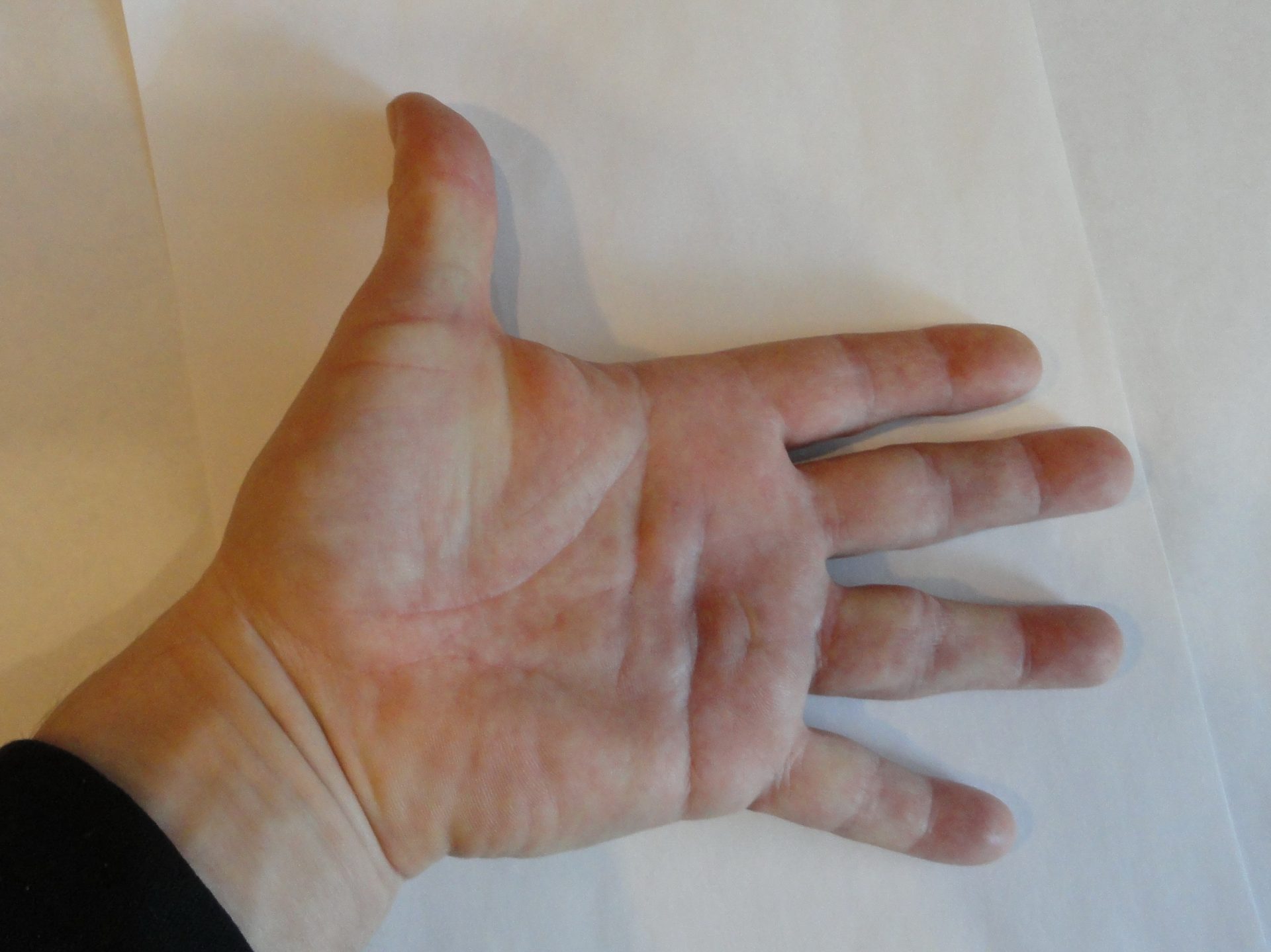
Vignette. A 24-year-old wide receiver presents to the training room one play after attempting to tackle a running back by grabbing the player’s jersey at the hip. He felt a sudden pop on the ulnar side of his right hand and now cannot bend the tip of his ring finger. On examination there is fusiform swelling of the ring finger with mild tenderness over the volar distal interphalangeal (DIP) joint. When the examiner stabilizes the proximal interphalangeal (PIP) joint in extension and asks the patient to flex the DIP, no motion is generated, although flexion at the PIP joint with the metacarpophalangeal (MCP) joint stabilized is preserved. Sensation is intact. Plain radiographs show no fracture.
What is the most likely diagnosis, what is the underlying anatomic structure that has failed, why is splinting alone the wrong answer here in contrast to the management of mallet finger, and what classification system governs the urgency of the surgical referral?
(Answer at the end of this chapter)
Section 1: Flexor Pulley System, Trigger Finger, and Dupuytren Contracture
Bottom line: the flexor pulley system holds the flexor digitorum superficialis (FDS) and flexor digitorum profundus (FDP) tendons against the phalanges with five annular pulleys (A1 to A5) and three cruciform pulleys (C1 to C3); A2 and A4 are biomechanically critical and must be preserved while A1 at the metacarpophalangeal (MCP) joint is the trigger finger site and can be safely released; trigger finger is treated stepwise with splinting, corticosteroid injection, and surgical A1 release; Dupuytren contracture is fibroproliferative disease of the palmar fascia producing progressive MCP-then-PIP flexion contractures in ring and small fingers of Northern European males with diabetes mellitus (DM), alcohol use, or epilepsy, treated with collagenase, needle aponeurotomy, or surgical fasciectomy when MCP contracture exceeds 30 degrees or any fixed PIP flexion is present.
The flexor pulley system is a series of fibrous bands that hold the flexor tendons against the phalanges so that they glide during finger flexion. Without these pulleys the tendons would bowstring away from bone. There are five annular pulleys (A1 to A5) and three cruciform pulleys (C1 to C3). The two most critical are A2 (over the proximal phalanx) and A4 (over the middle phalanx). Loss of either produces bowstringing and functional impairment, so A2 and A4 must be preserved, while A1 can be safely released.
The Camper chiasm is a critical anatomic feature inside this system. The FDS tendon splits into two slips at the proximal phalanx, the FDP passes through the gap, and the FDS slips reunite deep to the FDP and insert on the middle phalanx. The FDS flexes the PIP joint and the FDP flexes the DIP joint. An isolated FDP laceration eliminates DIP flexion but preserves PIP flexion. An isolated FDS laceration may not be obvious because the FDP can still flex the entire finger; the dedicated FDS test (block all other digits in extension and ask the patient to flex only the test finger at the PIP joint) identifies it.
Trigger finger, clinically called stenosing tenosynovitis, occurs at the A1 pulley at the MCP joint. Repetitive friction produces inflammation and thickening of the tendon sheath. A nodular swelling develops on the tendon, and when the nodule passes beneath the narrowed A1 pulley during flexion it catches and locks. The patient must passively force the finger into extension. A palpable nodule is felt at the MCP joint on the palmar side. The ring finger and thumb are most commonly affected. Risk factors include DM, rheumatoid arthritis (RA), repetitive gripping, and hypothyroidism; DM is the most testable association.
Initial management is splinting the MCP joint in extension. If splinting fails, corticosteroid injection into the tendon sheath is effective in approximately 60 to 70 percent of nondiabetic patients. Refractory or recurrent cases are treated with surgical release of the A1 pulley.
Dupuytren contracture is a fibroproliferative disorder of the palmar fascia. Nodular thickening progresses to form cords that extend from the palm into the fingers, contracting over time and pulling the fingers into MCP flexion first, then PIP. The ring finger is most commonly affected, followed by the small finger; the index and thumb are rarely involved. PIP contractures are more functionally limiting and harder to correct surgically.
The classic patient is a male of Northern European descent (“Viking disease”). Additional risk factors include DM, alcohol use, epilepsy, and family history. Patients with bilateral hand involvement, early onset, or involvement of the feet (Ledderhose disease) or penis (Peyronie disease) are said to have Dupuytren diathesis. The Hueston tabletop test is the bedside check: if the patient cannot lay the palm flat with all fingers fully extended, intervention is warranted. Garrod nodes (knuckle pads on the dorsum of the PIP joints) flag the diathesis pattern. Intervention thresholds: MCP contracture ≥ 30 degrees or any fixed PIP flexion.
Three treatment options exist. Collagenase clostridium histolyticum injection (Xiaflex) is injected directly into the cord; the enzyme dissolves collagen over 24 hours, after which the clinician manually ruptures the weakened cord. Needle aponeurotomy uses a hypodermic needle to perforate the cord, followed by manual rupture. Surgical fasciectomy is open excision of the diseased palmar fascia; it has the lowest recurrence rate but the highest morbidity.

Source: Andrew Pertsev, Wikimedia Commons, CC BY-SA 4.0. Clinical photograph of Dupuytren contracture showing characteristic palmar cord and finger flexion contracture from fibroproliferative thickening of the palmar fascia.
High Yield — Pulley system, trigger finger, and Dupuytren contracture
- A2 (proximal phalanx) and A4 (middle phalanx) are biomechanically critical: preserve (loss = bowstringing). A1 (MCP joint) = trigger finger site: safely released.
- Camper chiasm = FDS splits to let FDP pass through.
- Trigger finger = stenosing tenosynovitis at A1; locking in flexion + palpable MCP nodule; DM / RA risk factors. Ladder: MCP extension splinting, corticosteroid injection, surgical A1 release.
- Dupuytren contracture = palmar fascia fibroproliferation; ring > small finger; MCP first then PIP; male, Northern European, DM, alcohol, epilepsy, family history; Hueston tabletop test, Garrod nodes, Ledderhose (feet), Peyronie (penis) = diathesis.
- Dupuytren treatment: collagenase (Xiaflex), needle aponeurotomy, surgical fasciectomy. Thresholds: MCP ≥ 30 degrees or any fixed PIP contracture.
Mnemonic — “Two and four, hold the floor”
A2 over the proximal phalanx and A4 over the middle phalanx are the load-bearing pulleys. Lose them and the tendons bowstring. A1 at the MCP is the one that gets thick and locks; it can be released safely.
The A2 pulley over the proximal phalanx and the A4 pulley over the middle phalanx, those are the biomechanically non-negotiable ones. Losing either one of those basically cripples the finger. Because unlike A2 and A4, the A1 pulley is not biomechanically critical for preventing bowstringing. You release it, the nodule passes freely, and the finger functions normally.
— MSK-07-a podcast, ~00:38