Ultrasound · Foot
Peroneal Tendon Sheath Injection
ankleperonealretinaculumbrevislongus
Treats
- Peroneal tenosynovitis (primary indication — inflammation of the shared tendon sheath)
- Peroneal tendinopathy (peroneus longus/brevis)
Contraindications
- Active local skin infection at entry site (absolute)
- Suspected septic tenosynovitis (absolute)
- Known allergy to local anesthetic, steroid, or antiseptic prep
- Therapeutic anticoagulation — review case-by-case
- Uncontrolled diabetes (relative)
- High-grade peroneus brevis longitudinal split or tear (relative — favor non-particulate steroid or alternative therapy)
Pre-procedure checklist
- Confirm patient identity (two identifiers)
- Confirm procedure, side, and site
- Review allergies and anticoagulation
- Confirm informed consent
- REQUIRED pre-scan: map the sural nerve (most common iatrogenic injury at this site)
- Identify peroneus brevis for longitudinal split (changes steroid choice)
- Time-out immediately before needle entry
Positioning
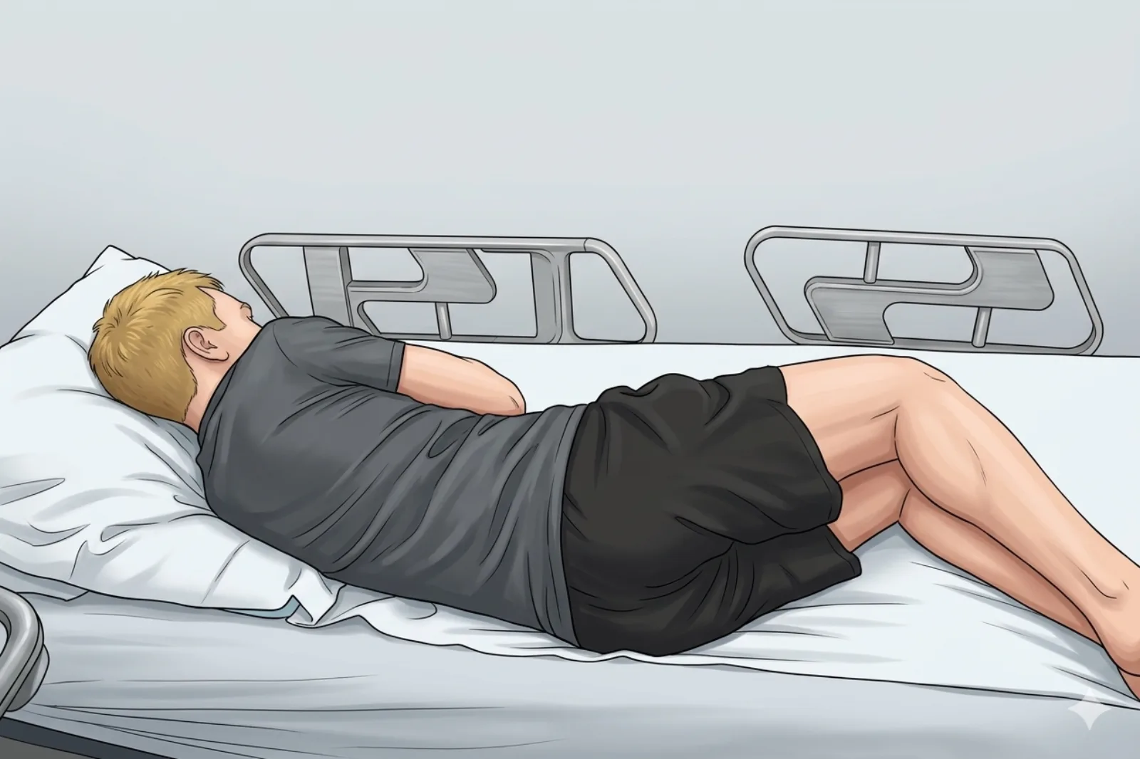
- Side-lying, symptomatic ankle up; foot in slight plantarflexion and inversion to relax the superior peroneal retinaculum and open the retromalleolar sulcus
Probe
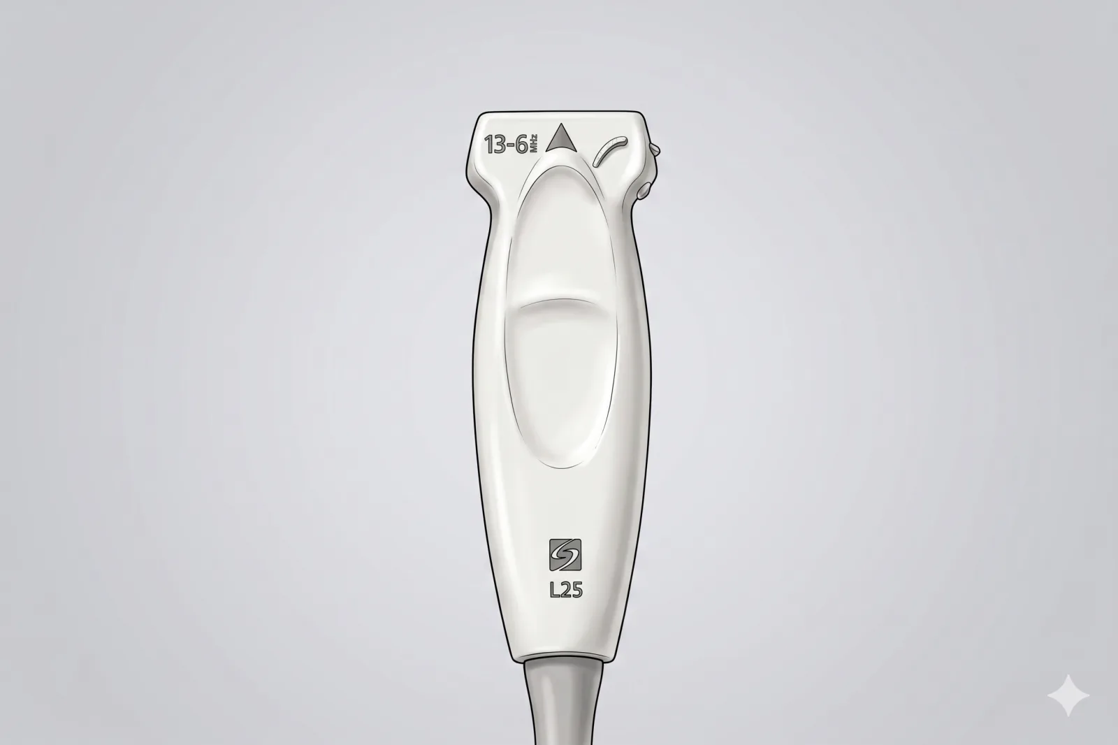
- High-frequency linear probe (6-13 MHz) (short footprint ideal)
Landmarking — find the view
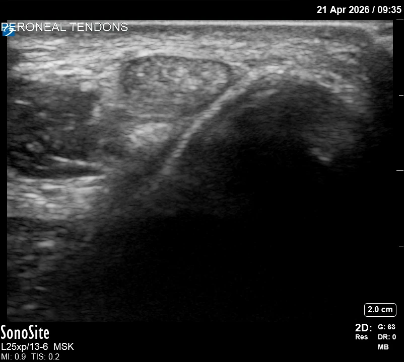
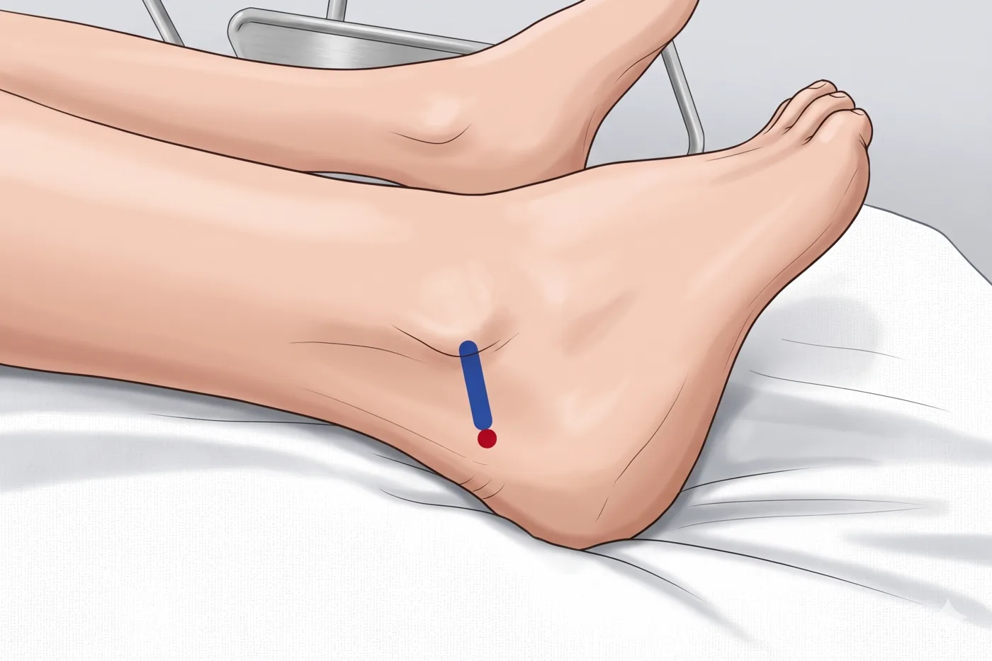
- Probe over posterior aspect of lateral malleolus → identify peroneus longus and brevis tendons sharing a sheath → see superior peroneal retinaculum overlying
The correct ultrasound view
- Two tendons in shared anechoic rim (sheath) behind the fibula
Needle corridor
- Anterior → posterior under retinaculum (above) into sheath around tendons (below)
Avoid
- Sural nerve — runs posterior to the peroneal tendons in the same fascial plane and is the most common iatrogenic injury at this site. Map it with US BEFORE every injection. Do not proceed if the nerve crosses the planned path.
- Small saphenous vein — accompanies the sural nerve posteriorly; identify with Doppler alongside the nerve survey
- Intratendinous steroid — particularly dangerous if peroneus brevis longitudinal split is present (rupture risk)
- Superficial fibular (peroneal) nerve — branches cross the anterolateral ankle near the skin entry; identify before needle entry.
Steps
- 1In-plane into sheath; surround both tendons with injectate.
- 2If injectate fails to spread freely around both tendons, WITHDRAW and reposition — do not increase injection pressure (failure to spread may indicate intratendinous placement).
- 3STOP IMMEDIATELY if the patient reports electric, shooting, or paresthetic sensations along the lateral foot or sural distribution.
Tips
- Short needle gives fine control; low volumes suffice.
- Limit to ≤ 2-3 injections per tendon per lifetime.
- If color Doppler shows a vessel in the planned path, abandon and re-plan.
Resident pearls
- Approach anterior-to-posterior under the superior peroneal retinaculum.
- Ensure injectate surrounds both the longus and brevis tendons in their shared sheath.
- Always survey for the sural nerve posteriorly before EVERY injection.
- Foot in slight plantarflexion + inversion relaxes the SPR and opens the working space.
Supplies
- 5 mL Syringe
- 25G 1.5" Needle
- 18G Drawing Needle
- 1% Lidocaine (1-2 mL)
- Dexamethasone 4-8 mg (PREFERRED — non-particulate, lower rupture risk near a split tendon) OR triamcinolone 20-40 mg
- Alcohol Swabs (vial tops)
- Betadine/Chloraprep
- Sterile Gloves
- Sterile 4x4 Gauze Pads
- Sterile Towels & Drapes
- Adhesive Bandage (Band-Aid)
- Washcloth/Towel (gel cleanup)
- Needle Disposal Safety Cap
- Sterile probe cover & gel
Post-procedure
- Apply a clean dressing; remove after 24 hours.
- Local anesthetic relief for 1-4 hours; steroid begins 24-72 hours.
- Steroid 'flare' in 2–10% — manage with ice and acetaminophen.
- Rest the ankle 24-48 hours; avoid heavy loading or running for 1 week.
- Return immediately for: spreading redness, swelling, fever, disproportionate pain, OR sudden 'pop' or weakness with push-off (concern for tendon rupture).
- Diabetics: expect 50-100 mg/dL glucose rise for 3-5 days.
- Maximum 2-3 injections per tendon lifetime.