Ultrasound · Foot
Tibiotalar (Ankle) Joint Injection
ankletibiotalardorsalis pedisdeep peroneal
Treats
- Ankle osteoarthritis
- Synovitis
- Diagnostic anesthetic
Contraindications
- Active local skin infection at entry site (absolute)
- Suspected septic joint (absolute — aspirate first, do not inject steroid)
- Bacteremia (absolute)
- Known allergy to local anesthetic, steroid, or antiseptic prep
- Therapeutic anticoagulation — review case-by-case
- Uncontrolled diabetes (relative)
- Ankle prosthesis at the target site (relative)
Pre-procedure checklist
- Confirm patient identity (two identifiers)
- Confirm procedure, side, and site — mark with patient awake
- Review allergies and anticoagulation
- Confirm informed consent
- Pre-scan: identify TA, EHL, EDL tendons; Doppler the deep peroneal nerve and dorsalis pedis artery (lateral to EHL); plan medial-to-EHL entry
- Time-out immediately before needle entry
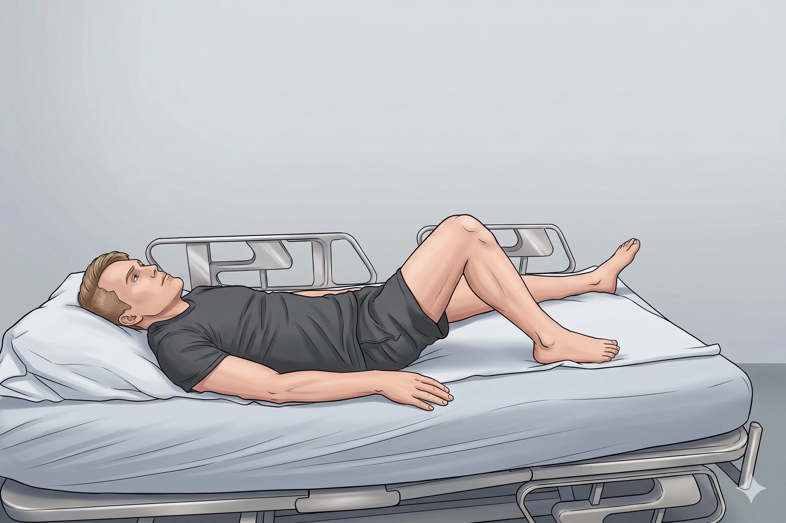
Positioning
- Supine; leg extended; ankle in slight plantarflexion
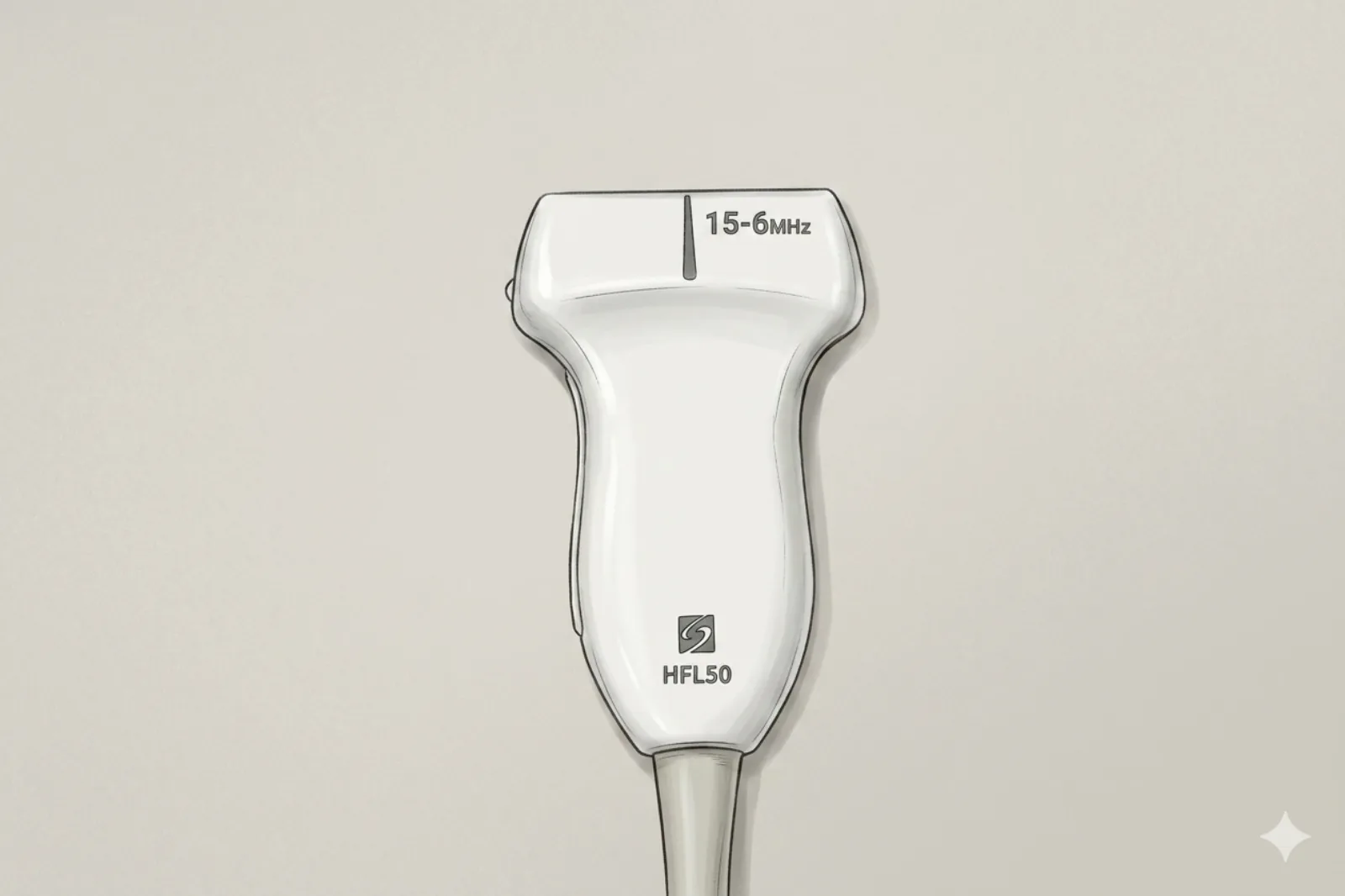
Probe
- High-frequency linear probe (6-13 MHz)
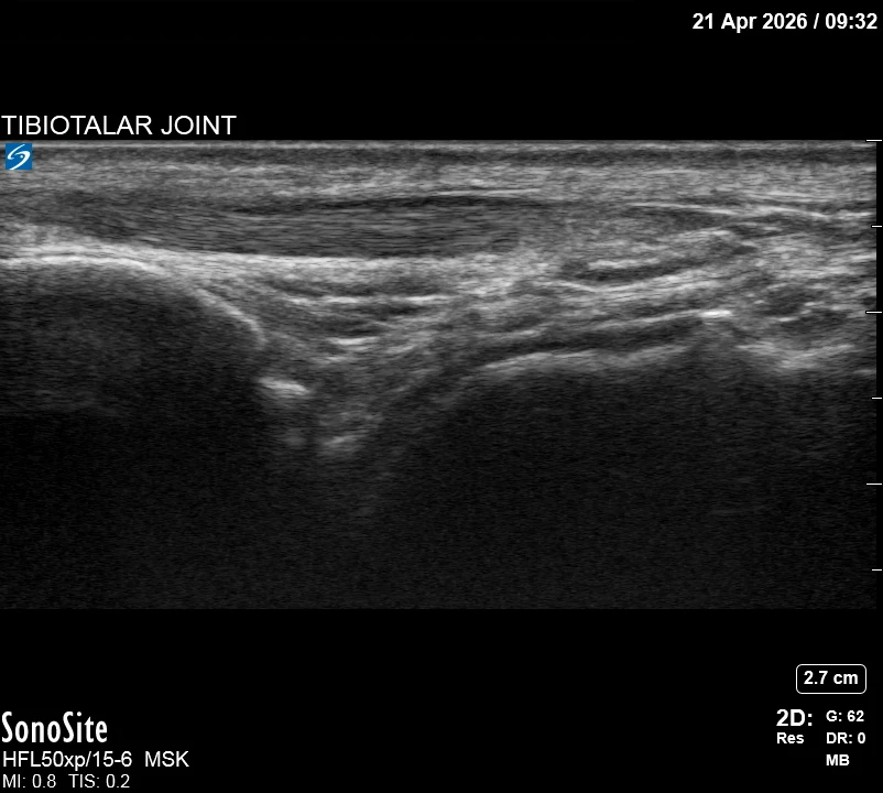
Landmarking — find the view
- Visual Cue: The 'Talar Dome Profile' — identify the smooth convex curve of the talus.
- Short-Axis Scan: identify the anterior tendons (TA, EHL, EDL).
- Doppler Survey: Map the 'No-Fly Zone' — deep peroneal nerve and dorsalis pedis artery (lying between EHL and EDL on the lateral side).
- Identify the joint capsule as a distinct dark plane just above the talar cartilage.
The correct ultrasound view
- Identify the talar dome (curved line) with distinct dark capsular line above; tendons are superficial and out of path
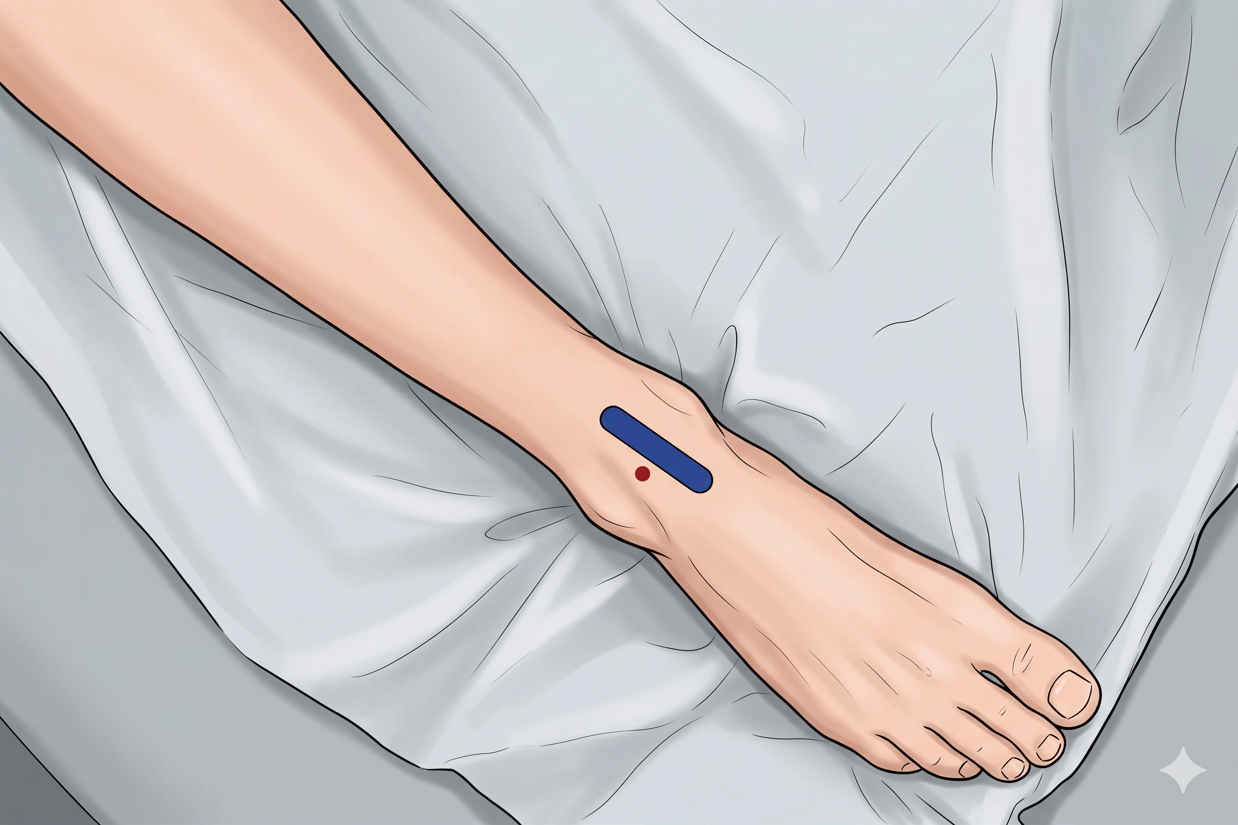
Needle corridor
- Anteromedial entry BETWEEN the tibialis anterior tendon (medially) and the extensor hallucis longus tendon (laterally), targeting the medial gutter of the tibiotalar joint. Keep the needle medial to the EHL at all times — the dorsalis pedis artery and deep peroneal nerve lie lateral to EHL. Do not advance toward the lateral compartment.
Avoid
- Deep peroneal nerve and dorsalis pedis artery (lateral to EHL — stay medial of them throughout)
- Intratendinous injection of TA, EHL, or EDL
Steps
- 1In-plane entry between the tibialis anterior and EHL tendons (medial to EHL); hug capsule superficial to cartilage.
- 2Inject as indicated; confirm intra-articular distension.
- 3STOP IMMEDIATELY if the patient reports electric, shooting, or paresthetic sensations into the foot.
Tips
- Small tilts separate capsule from cartilage and improve needle visualization.
- Limit corticosteroid to ≤ 3 per joint per year.
- If color Doppler shows the DPA in the planned path, abandon and re-plan.
Resident pearls
- Enter between the tibialis anterior and EHL tendons (medial to EHL). The DPA/DPN bundle lies LATERAL to EHL — stay medial to EHL throughout.
- The target is the space between the joint capsule and the talar hyaline cartilage.
Supplies
- For injection only: 25G 1.5" needle
- For aspiration ± injection (suspected effusion, synovitis workup, septic rule-out): 20G or 22G 1.5" needle
- 5 mL Syringe
- 18G Drawing Needle
- 1% Lidocaine (3-5 mL)
- Triamcinolone 40 mg OR Dexamethasone 4-10 mg
- Alcohol Swabs (vial tops)
- Betadine/Chloraprep
- Sterile Gloves
- Sterile 4x4 Gauze Pads
- Sterile Towels & Drapes
- Adhesive Bandage (Band-Aid)
- Washcloth/Towel (gel cleanup)
- Needle Disposal Safety Cap
- Sterile probe cover & gel
Post-procedure
- Apply a clean dressing; remove after 24 hours.
- Local anesthetic relief for 1-4 hours; steroid begins 24-72 hours.
- Steroid 'flare' in 2–10% — manage with ice and acetaminophen.
- Rest the ankle 24-48 hours; resume normal activity as tolerated thereafter.
- Return immediately for: spreading redness, swelling, fever, disproportionate pain, OR new foot numbness or weakness.
- Diabetics: expect 50-100 mg/dL glucose rise for 3-5 days.
- Maximum 3 injections per joint per year.