Ultrasound · Elbow
Common Extensor Tendon Origin Injection (Lateral Epicondyle)
elbowtennis elbowcet
Treats
- Lateral epicondylitis with tendon surface pain or degeneration
Contraindications
- Active local skin infection at entry site (absolute)
- Known allergy to local anesthetic, steroid, or antiseptic prep
- Therapeutic anticoagulation — review case-by-case
- Uncontrolled diabetes (relative)
- High-grade tendon tear (relative — favor regenerative injectate over steroid)
Pre-procedure checklist
- Confirm patient identity (two identifiers)
- Confirm procedure, side, and site — mark with patient awake
- Review allergies and anticoagulation
- Confirm informed consent
- Pre-scan to map radial nerve and radial recurrent artery
- Time-out immediately before needle entry
Positioning
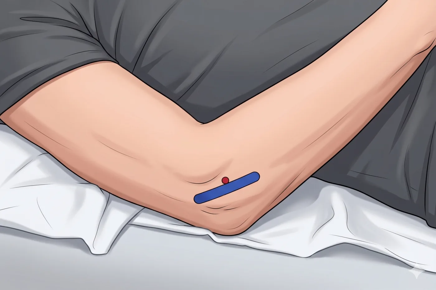
- Supine; elbow 20-40° flexed; hand on abdomen
.webp)
Probe
- High-frequency linear probe (6-13 MHz)
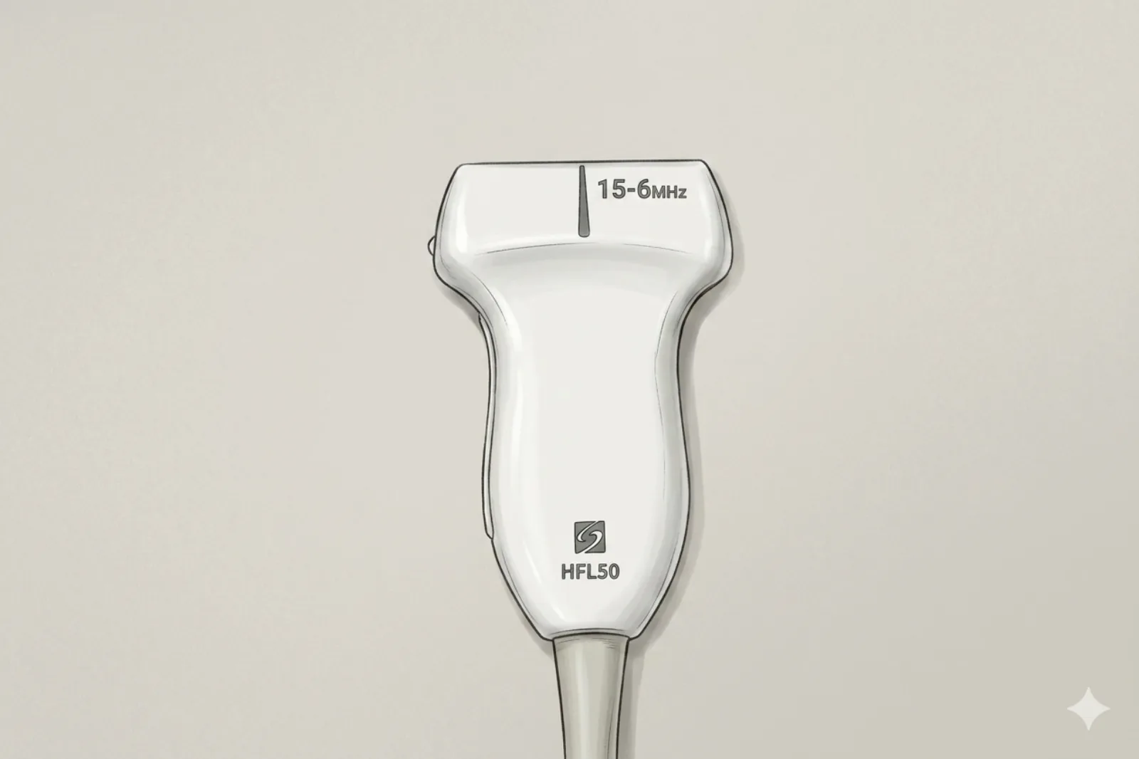
Landmarking — find the view
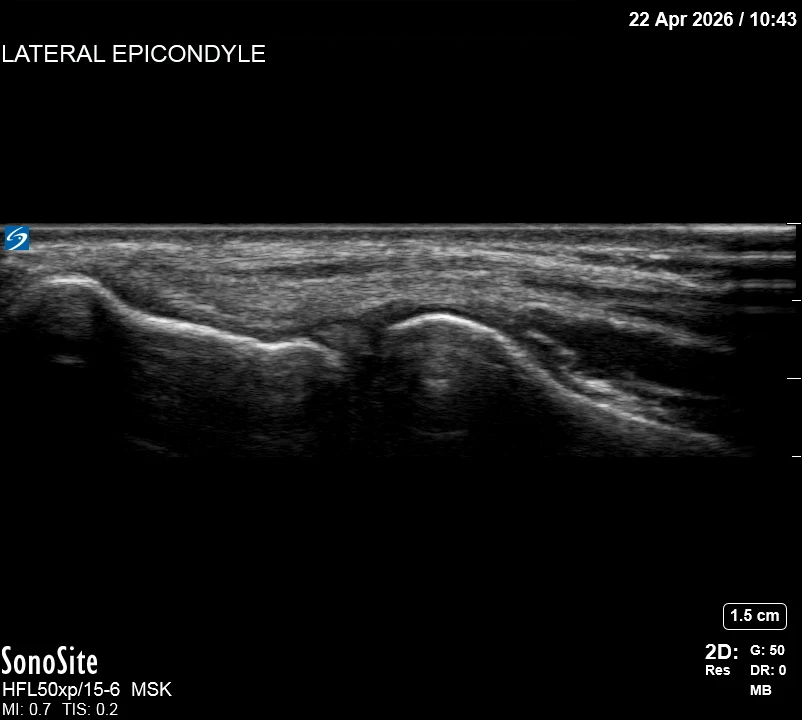
- Visual Cue: The “Fibrillar Footprint” — Identify the bright, organized fibers of the CET attaching to the lateral epicondyle.
- Slide the probe slightly anteriorly to identify the landmark bony contour of the radial head.
- Check for any hypoechoic “tears” or calcifications within the footprint.
- Map the radial nerve and the radial recurrent artery before choosing your needle path.
The correct ultrasound view
- Crisp fibrillar tendon superficial to thin bright ligament band
Needle corridor
- Distal → proximal path between skin/fat (above) and tendon surface (below) for peritendinous steroid
Avoid
- Radial nerve (superficial branch runs anterior to the radial head at this level — identify before entry)
- Radial recurrent artery
- Lateral ulnar collateral ligament (LUCL) — originates at the lateral epicondyle; deep injection into or through the LUCL risks posterolateral rotatory instability (PLRI). Keep needle superficial to the tendon surface.
- Intratendinous steroid
Steps
- 1Peritendinous steroid: deposit aliquots in the plane BETWEEN the fascial/fat layer and the tendon surface — NOT within the fibrillar tendon itself.
- 2For regenerative injectates (PRP, percutaneous tenotomy), target intratendinous diseased zones — this is a different technique and not for steroid.
- 3STOP IMMEDIATELY if the patient reports electric, shooting, or paresthetic sensations.
Tips
- Limit corticosteroid here to 1-2 injections lifetime — and consider non-steroid alternatives first (see pearls).
- If color Doppler shows the radial recurrent artery in the planned path, abandon and re-plan.
Resident pearls
- Corticosteroid for tendinopathy provides short-term relief but is associated with WORSE long-term outcomes in published RCTs (Coombes BK et al. JAMA 2013;309:461). Consider PRP, percutaneous tenotomy, or peritendinous saline as alternatives. If steroid is used, single low-dose injection only.
- The radial collateral ligament is deep — forming the deep 30-50% of the common-origin footprint.
- Avoid placing corticosteroid directly into the tendon fibers to prevent future rupture.
Supplies
- 3 mL Syringe
- 25G or 27G 1" Needle
- 18G Drawing Needle
- 1% Lidocaine (1-2 mL)
- Triamcinolone 20 mg OR Dexamethasone 4 mg (single low-dose, peritendinous only)
- Alcohol Swabs (vial tops)
- Betadine/Chloraprep
- Sterile Gloves
- Sterile 4x4 Gauze Pads
- Sterile Towels & Drapes
- Adhesive Bandage (Band-Aid)
- Washcloth/Towel (gel cleanup)
- Needle Disposal Safety Cap
- Sterile probe cover & gel
Post-procedure
- Apply a clean dressing; remove after 24 hours.
- Local anesthetic relief for 1-4 hours; steroid begins 24-72 hours.
- Steroid 'flare' in 2–10% — manage with ice and acetaminophen.
- Rest the forearm 24-48 hours; avoid heavy gripping for 1 week.
- Return immediately for: spreading redness, swelling, fever, or disproportionate pain.
- Diabetics: expect 50-100 mg/dL glucose rise for 3-5 days.
- Maximum 1-2 steroid injections at this site lifetime — recurrent dosing worsens outcomes.