Ultrasound · Elbow
Radiocapitellar Joint Injection (Lateral Elbow)
elbowradiocapitellararthritissynovitis
Treats
- Elbow arthritis
- Lateral elbow joint pain
- Synovitis/effusion
- Diagnostic anesthetic
Contraindications
- Active local skin infection at entry site (absolute)
- Suspected septic joint (absolute — aspirate first)
- Bacteremia (absolute)
- Known allergy to local anesthetic, steroid, or antiseptic prep
- Therapeutic anticoagulation — review case-by-case
- Uncontrolled diabetes (relative)
Pre-procedure checklist
- Confirm patient identity (two identifiers)
- Confirm procedure, side, and site — mark with patient awake
- Review allergies and anticoagulation
- Confirm informed consent
- Pre-scan to identify and trace the radial nerve and PIN around the radial neck
- Time-out immediately before needle entry
Positioning
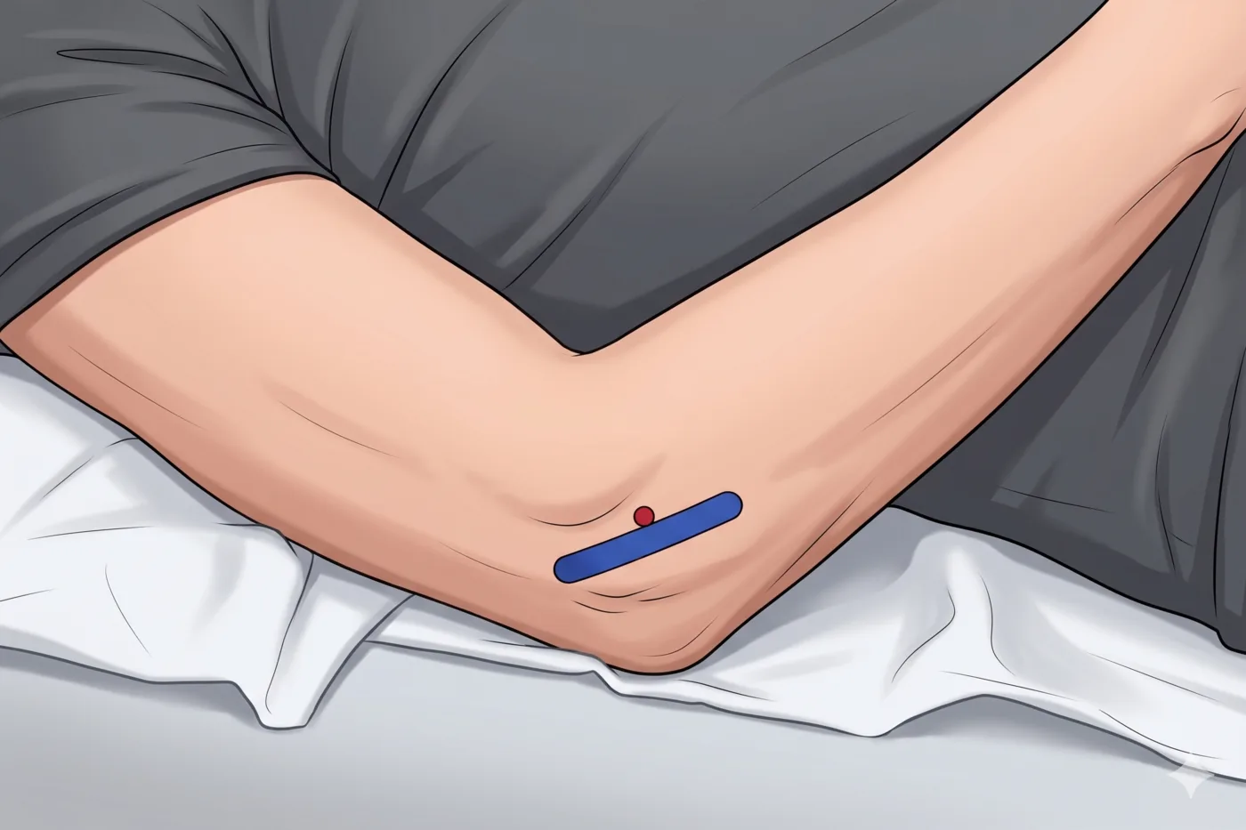
- Supine; elbow flexed 20-40°; hand on abdomen; small pillow under elbow
.webp)
Probe
- High-frequency linear probe (6-13 MHz)
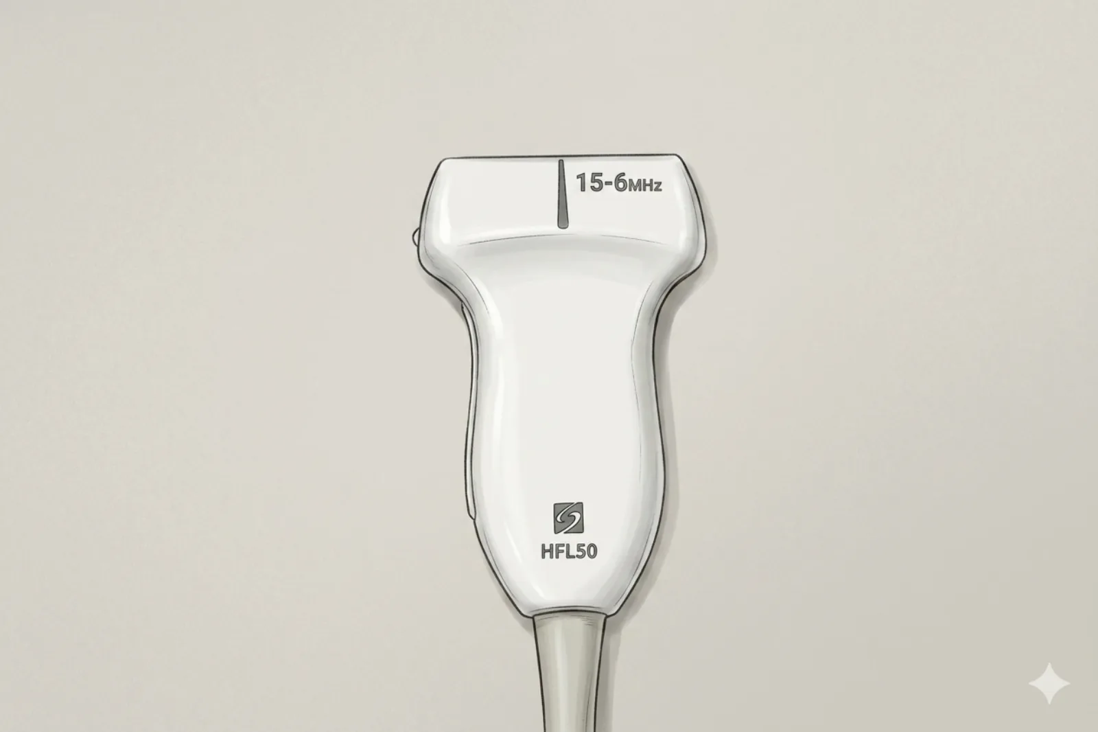
Landmarking — find the view
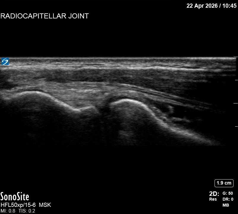
- Find lateral epicondyle → slide distally to radial head → tilt anteriorly to view capitellum → identify wedge-shaped joint space
The correct ultrasound view
- Clear radial head–capitellum interface with a thin dark joint space
Needle corridor
- Preferred: in-plane lateral approach with the needle parallel to the probe long-axis, advancing into the joint cleft under continuous visualization.
Avoid
- Radial nerve and its posterior interosseous nerve (PIN) branch — the PIN wraps around the radial neck through the supinator (arcade of Frohse) immediately distal to the radiocapitellar joint. Identify and trace before needle entry.
- Radial recurrent vessels
- Lateral ulnar collateral ligament (LUCL) — located at the posteroinferior aspect of the lateral joint; repeated needle trauma or intralesional injection risks posterolateral rotatory instability (PLRI)
Steps
- 1Preferred: in-plane lateral approach into the joint cleft. Out-of-plane walk-down is an alternative for clinicians comfortable with the technique.
- 2Test inject; inject 2-3 mL total, stopping if capsular back-pressure is felt.
- 3STOP IMMEDIATELY if the patient reports electric, shooting, or paresthetic sensations.
Tips
- Walk-down (when used) intentionally uses gentle periosteal contact on the capitellum to localize the joint cleft — this is different from advancing blindly through soft tissue. Never force against articular cartilage.
- Pronate/supinate the forearm to confirm the radial head moves while the lateral epicondyle stays stationary.
- Limit corticosteroid to ≤ 3 per joint per year.
- If color Doppler shows a vessel directly in the planned path, abandon and re-plan.
Resident pearls
- Visual Orientation: Think of the joint as a “cleft” between two hills. The capitellum is the rounded hill, and the radial head is the flatter hill.
- Native radiocapitellar joint capacity is ~1-3 mL — total injectate ≤ 3 mL. Over-distension risks capsular rupture and steroid extravasation toward the PIN/radial nerve.
Supplies
- 5 mL Syringe
- 22G or 25G 1.5" Needle
- 18G Drawing Needle
- 1% Lidocaine (1-2 mL)
- Triamcinolone 20-40 mg OR Dexamethasone 4-10 mg
- Alcohol Swabs (vial tops)
- Betadine/Chloraprep
- Sterile Gloves
- Sterile 4x4 Gauze Pads
- Sterile Towels & Drapes
- Adhesive Bandage (Band-Aid)
- Washcloth/Towel (gel cleanup)
- Needle Disposal Safety Cap
- Sterile probe cover & gel
Post-procedure
- Apply a clean dressing; remove after 24 hours.
- Local anesthetic relief for 1-4 hours; steroid begins 24-72 hours.
- Steroid 'flare' in 2–10% — manage with ice and acetaminophen.
- Rest the elbow 24-48 hours.
- Return immediately for: spreading redness, swelling, fever, or disproportionate pain.
- Diabetics: expect 50-100 mg/dL glucose rise for 3-5 days.
- Maximum 3 injections per joint per year.