Ultrasound · Foot
Morton's Neuroma Injection (Dorsal Approach)
footmorton's neuromametatarsalweb space
Treats
- Interdigital neuralgia (Morton's neuroma)
- Intermetatarsal bursitis
Contraindications
- Active local skin infection at entry site (absolute)
- Known allergy to local anesthetic, steroid, or antiseptic prep
- Therapeutic anticoagulation — review case-by-case
- Uncontrolled diabetes (relative)
Pre-procedure checklist
- Confirm patient identity (two identifiers)
- Confirm procedure, side, and TARGET WEB SPACE — mark with patient awake
- Review allergies and anticoagulation
- Confirm informed consent
- Pre-scan: identify the neuroma in the target web space; perform Mulder's maneuver to confirm; identify the deep transverse intermetatarsal ligament; Doppler deep plantar metatarsal vessels
- Time-out immediately before needle entry
Positioning
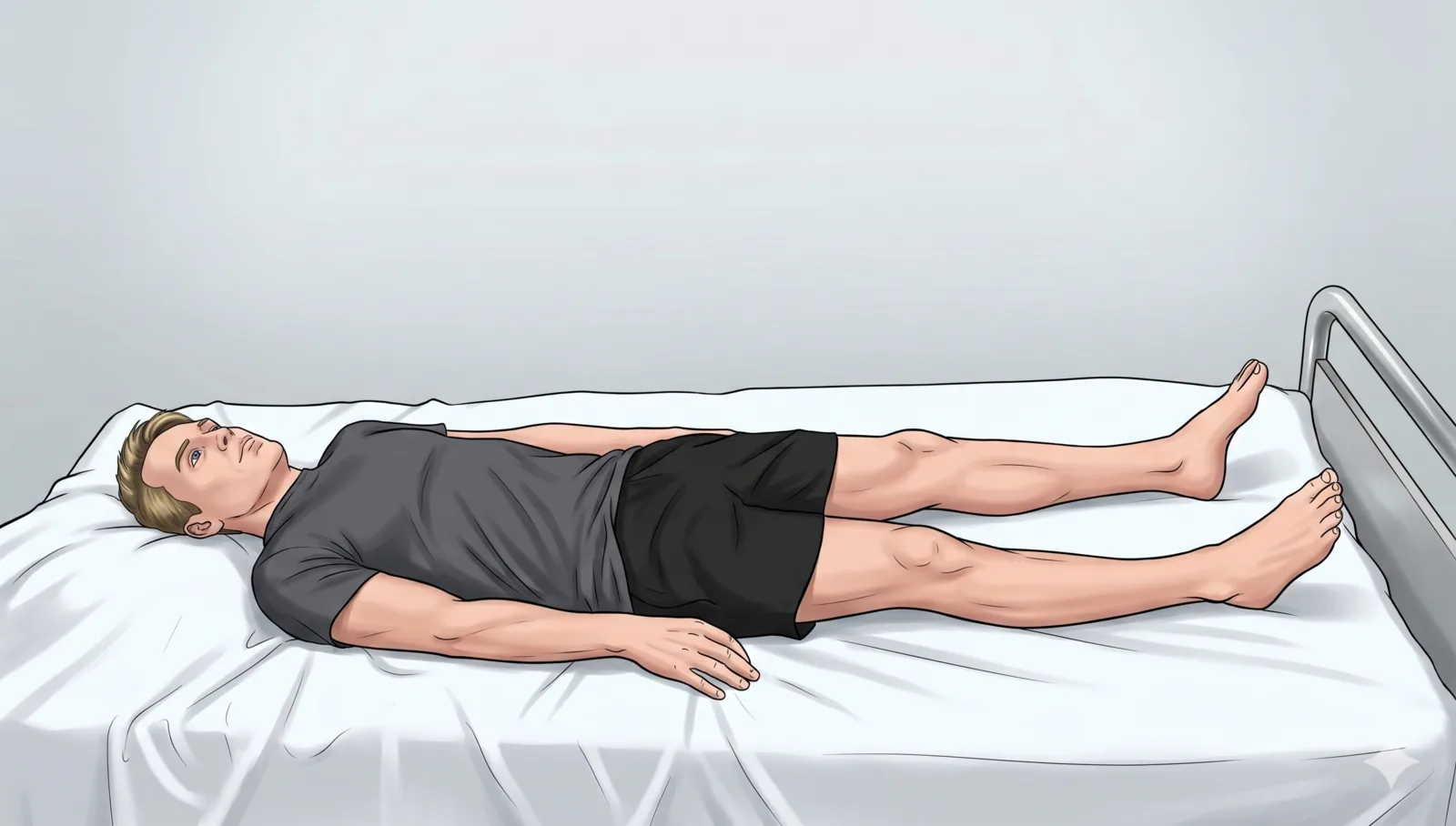
- Supine; foot resting on table; toes slightly flexed
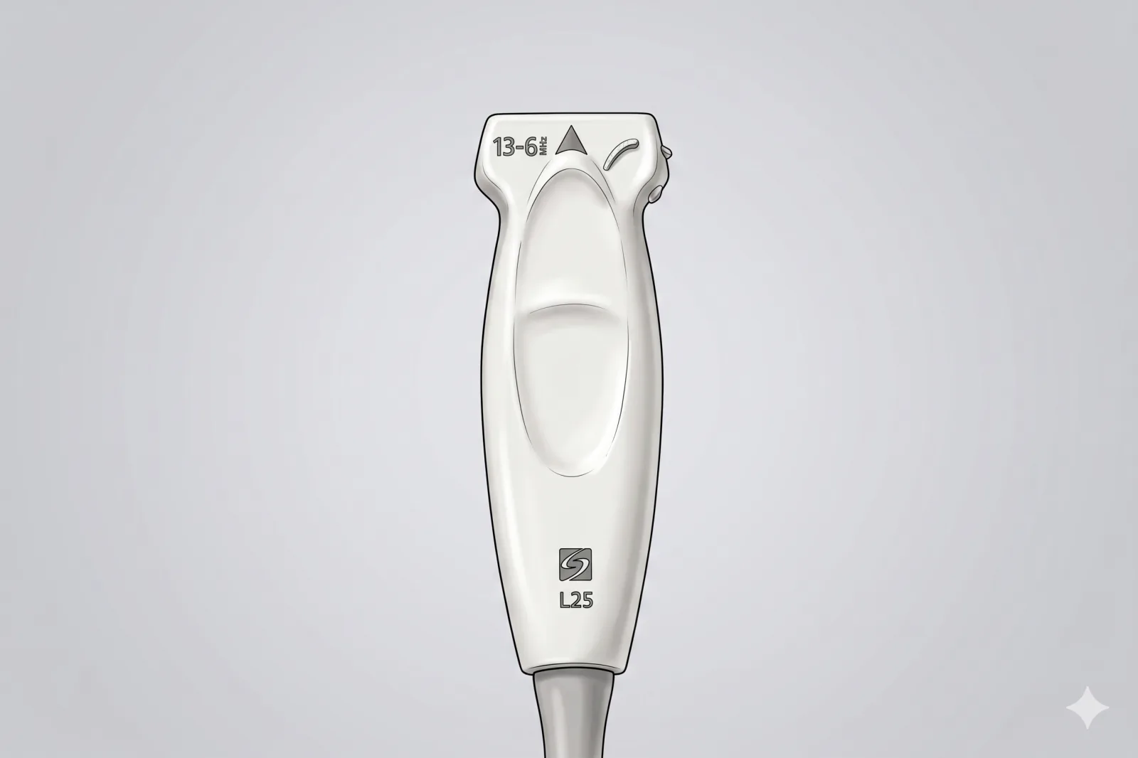
Probe
- High-frequency linear probe (6-13 MHz)
Landmarking — find the view
- Mulder's Maneuver: Squeeze the forefoot transversely (medial-lateral compression of the metatarsal heads) while simultaneously applying plantar pressure on the suspect interspace. Watch in real-time for the hypoechoic neuroma to 'pop' dorsally between the metatarsal heads — this is the sonographic Mulder's click and confirms target identification.
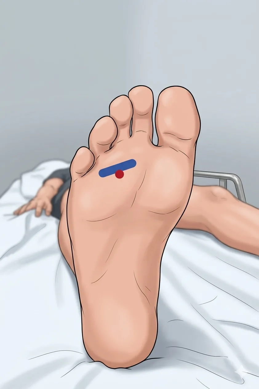
- Transverse Scan: Identify the target web space (usually 2nd-3rd or 3rd-4th).
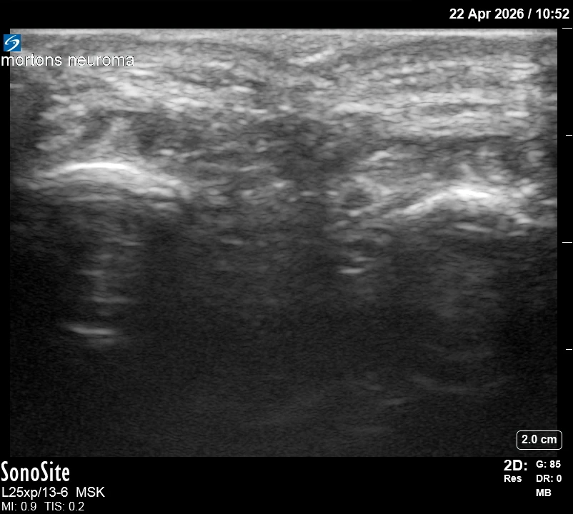
- Identify the hypoechoic 'dumbbell' or round mass as it 'pops' dorsally during compression.
- Identify and avoid the intermetatarsal bursa, which often sits just superficial to the neuroma.
The correct ultrasound view
- Hypoechoic mass in the intermetatarsal space; metatarsal heads on either side.
Needle corridor
- Dorsal in-plane approach directly into the intermetatarsal space, traversing the deep transverse intermetatarsal ligament to reach the neuroma plantar to it.
Avoid
- Deep plantar metatarsal vessels
- Adjacent (non-target) digital nerves in neighboring web spaces. Note: target IS the common plantar digital nerve — aim for PERI-NEURAL placement around the hypoechoic mass, not intraneural.
Steps
- 1Advance dorsal-to-plantar under real-time US.
- 2Feel for a subtle 'pop' as the needle traverses the deep transverse intermetatarsal ligament.
- 3Target the peri-neural space around the hypoechoic mass.
- 4Inject 0.5-1 mL total (lidocaine + steroid premixed); watch for fluid surrounding the neuroma.
- 5STOP IMMEDIATELY if the patient reports electric, shooting, or paresthetic sensations into the toes.
Tips
- Dorsal approach is safer and avoids painful plantar weight-bearing skin.
- Larger volumes risk spread to adjacent interspaces and plantar fat pad atrophy.
- Limit to 2-3 injections per site per year.
- If color Doppler shows a deep plantar vessel in the planned path, abandon and re-plan.
Resident pearls
- Mulder's = medial-lateral squeeze + plantar push. Don't rely on plantar-only pressure.
- The neuroma sits PLANTAR to the deep transverse intermetatarsal ligament. From a dorsal approach, feel for a subtle 'pop' through the ligament and confirm tip is deep to it. Stopping above the ligament deposits steroid in the intermetatarsal bursa instead of around the neuroma.
- Identify and avoid the intermetatarsal bursa, which may also be inflamed.
- Steroid in the plantar fat pad can cause atrophy — the dorsal approach minimizes this risk.
Supplies
- 3 mL Syringe
- 25G 1" or 1.5" Needle (use ≥ 25G if injecting triamcinolone — 27G can clog with particulate suspension)
- 18G Drawing Needle
- 1% Lidocaine (0.5 mL)
- Dexamethasone 4 mg (PREFERRED — non-particulate, no clog risk, lower fat-atrophy risk) OR triamcinolone 20 mg
- Alcohol Swabs (vial tops)
- Betadine/Chloraprep
- Sterile Gloves
- Sterile 4x4 Gauze Pads
- Sterile Towels & Drapes
- Adhesive Bandage (Band-Aid)
- Washcloth/Towel (gel cleanup)
- Needle Disposal Safety Cap
- Sterile probe cover & gel
Post-procedure
- Apply a clean dressing; remove after 24 hours.
- Local anesthetic relief for 1-4 hours; steroid begins 24-72 hours.
- Steroid 'flare' in 2–10% — manage with ice and acetaminophen.
- Post-injection neuritis: transient burning or aching in the toes for 24-72 hours is well-recognized after interdigital nerve injection — counsel the patient before the procedure.
- Rest the foot 24-48 hours; avoid tight footwear and wear cushioned shoes.
- Return immediately for: spreading redness, swelling, fever, disproportionate pain, OR new toe numbness or color change.
- Diabetics: expect 50-100 mg/dL glucose rise for 3-5 days.
- Maximum 2-3 injections per site per year.