Ultrasound · Foot
Plantar Fascia (Perifascial) Injection
footplantar fasciacalcaneusheelfat pad
Treats
- Plantar fasciitis (heel pain) requiring perifascial therapy
- NOT for plantar fibromatosis, retrocalcaneal bursitis, or tarsal tunnel syndrome — re-diagnose if US shows a mass lesion or tunnel-level pathology
Contraindications
- Active local skin infection at entry site (absolute)
- Known allergy to local anesthetic, steroid, or antiseptic prep
- Therapeutic anticoagulation — review case-by-case
- Uncontrolled diabetes (relative)
- Plantar fibromatosis or other mass lesion (re-diagnose, not for this procedure)
Pre-procedure checklist
- Confirm patient identity (two identifiers)
- Confirm procedure, side, and site
- Review allergies and anticoagulation
- Confirm informed consent — DISCUSS fat pad atrophy and fascial rupture risks
- Pre-scan: identify plantar fascia thickness, fat pad, calcaneus; map medial calcaneal nerve and Baxter's nerve; Doppler the medial NV bundle
- Time-out immediately before needle entry
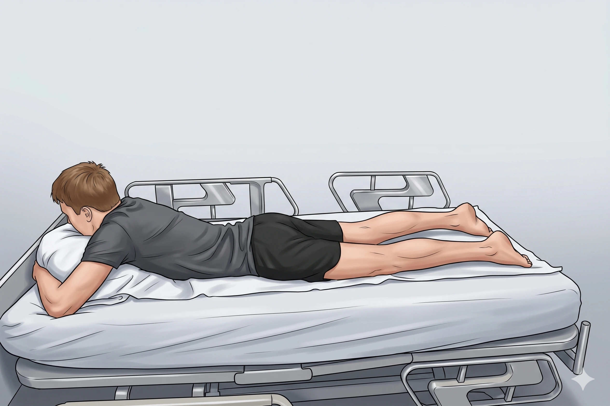
Positioning
- Prone or seated with the foot hanging off the table edge (preferred — provides direct plantar access and places the fascia under slight tension for better visualization). Supine with the foot flat on the table is an acceptable alternative for the medial approach but limits direct plantar heel access.
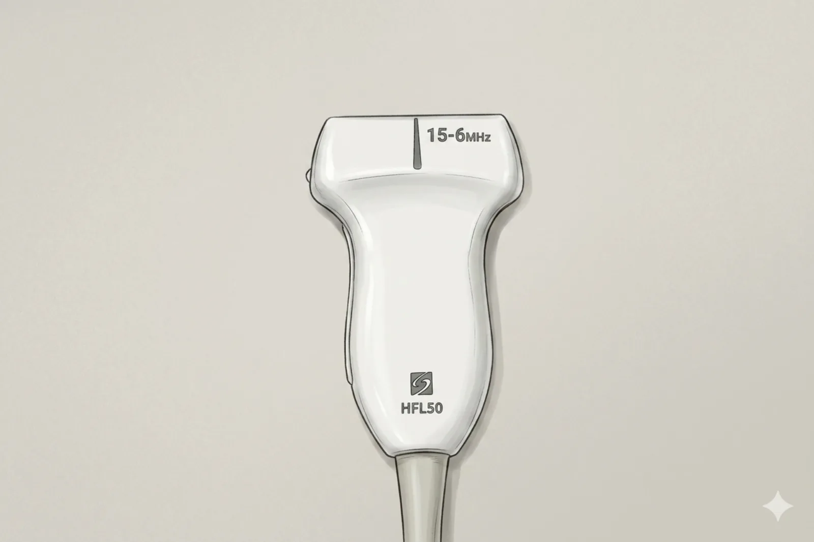
Probe
- High-frequency linear probe (6-13 MHz)
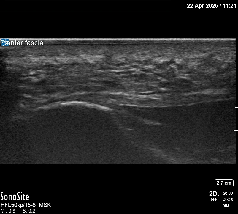
Landmarking — find the view
- Short-axis over plantar heel at calcaneal origin → identify bright plantar fascia on calcaneus → find thin dark plane between fascia and fat pad
The correct ultrasound view
- Bright fascia with thin anechoic plane separating it from fat pad
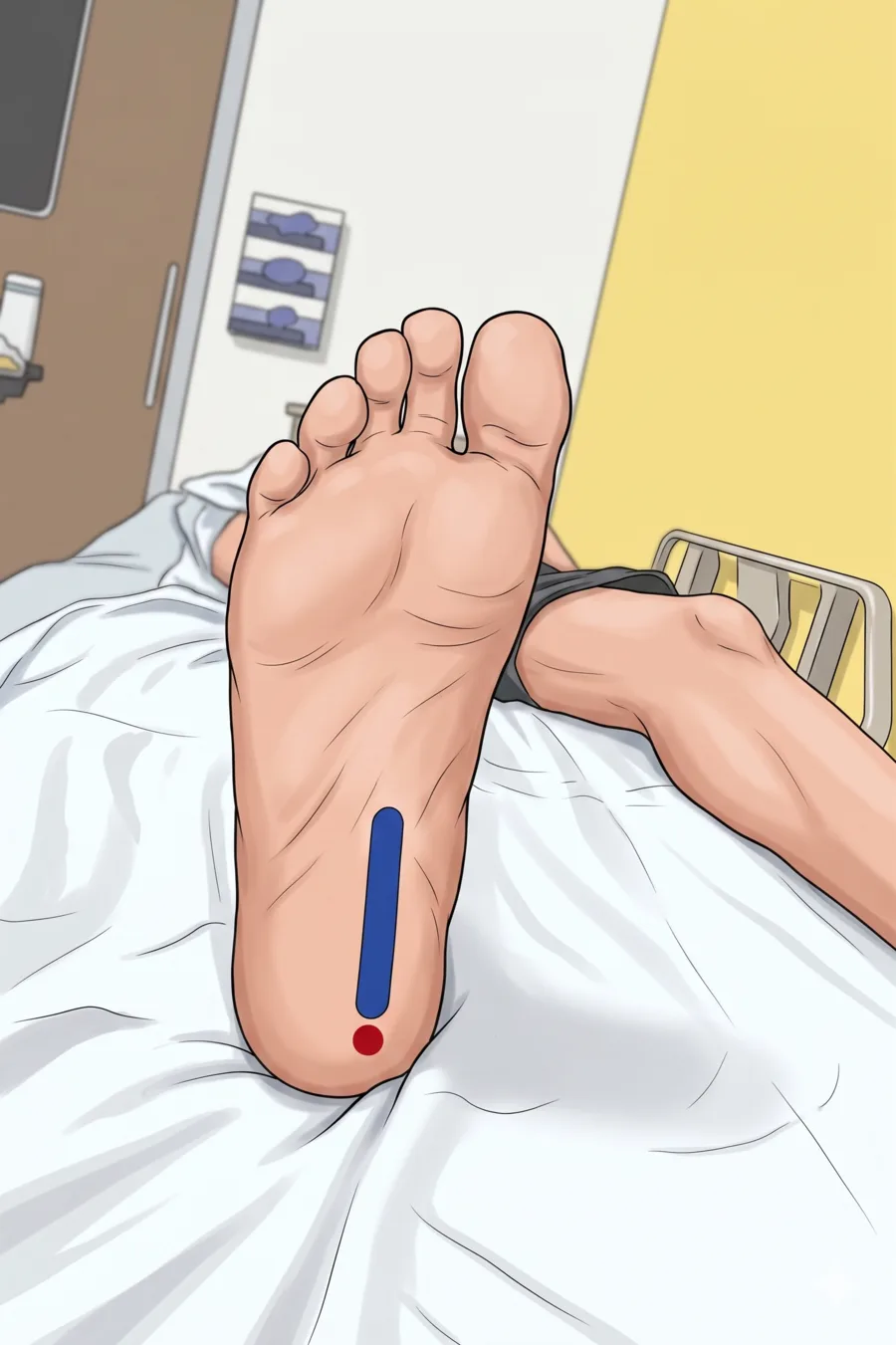
Needle corridor
- Medial → lateral within thin plane: fat pad (above), plantar fascia (below) — do not enter fascia
Avoid
- Intrafascial steroid (rupture risk)
- Injection into fat pad (atrophy risk)
- Medial calcaneal branch of the tibial nerve (superficial sensory to medial heel)
- Baxter's nerve (1st branch of lateral plantar n., between abductor hallucis and quadratus plantae at the depth of the fascia origin)
- Posterior tibial NV bundle (deeper in tarsal tunnel)
Steps
- 1In-plane into the perifascial plane; inject small volumes to expand plane without entering fascia.
- 2STOP IMMEDIATELY if the patient reports electric, shooting, or paresthetic sensations into the heel or sole.
- 3Advance the needle in-plane, medial to lateral, until the tip lies immediately deep to the fascia at its calcaneal origin.
- 4Inject slowly DEEP to the fascia — never into the heel fat pad; watch for anechoic spread along the fascial undersurface.
- 5Withdraw, apply pressure, and re-scan to confirm the spread pattern.
Tips
- Use smallest effective volume; total injectate ≤ 3 mL.
- Stop if resistance — correct plane opens smoothly. If you meet resistance and push harder, steroid can extravasate INTO the fascia (rupture risk).
- Limit to 1-2 steroid injections at this site lifetime (cumulative rupture and atrophy risk).
- If color Doppler shows the medial NV bundle in the planned path, abandon and re-plan.
Resident pearls
- The target is the hypoechoic plane between the fat pad (superficial) and fascia (deep).
- Avoid intrafascial or fat pad injection to prevent rupture or atrophy.
- Approach medial-to-lateral to avoid the medial neurovascular bundle.
- Watch out for Baxter's nerve entrapment as a separate cause of heel pain — distinct diagnosis requiring different treatment.
Supplies
- 5 mL Syringe
- 25G 1.5" Needle (default; 27G only with non-particulate steroid — triamcinolone can clog 27G)
- 18G Drawing Needle
- 1% Lidocaine (1-2 mL)
- Dexamethasone 4 mg (PREFERRED — lower atrophy/rupture risk) OR triamcinolone 20 mg (single low dose)
- Alcohol Swabs (vial tops)
- Betadine/Chloraprep
- Sterile Gloves
- Sterile 4x4 Gauze Pads
- Sterile Towels & Drapes
- Adhesive Bandage (Band-Aid)
- Washcloth/Towel (gel cleanup)
- Needle Disposal Safety Cap
- Sterile probe cover & gel
Post-procedure
- Apply a clean dressing; remove after 24 hours.
- Local anesthetic relief for 1-4 hours; steroid begins 24-72 hours.
- Steroid 'flare' in 2–10% — manage with ice and acetaminophen.
- Rest the heel 48-72 hours; avoid running and prolonged standing for 1 week.
- Return immediately for: spreading redness, swelling, fever, disproportionate pain, OR sudden tearing sensation in the heel (concern for fascial rupture).
- Diabetics: expect 50-100 mg/dL glucose rise for 3-5 days.
- Maximum 1-2 steroid injections at this site lifetime.