Ultrasound · Hand
First Carpometacarpal (CMC) Joint Injection (Thumb Base / Basal Joint)
handcmcthumb basebasal jointarthritis
Treats
- Thumb basal joint (1st CMC) arthritis or synovitis
Contraindications
- Active local skin infection at entry site (absolute)
- Suspected septic joint (absolute — aspirate first)
- Known allergy to local anesthetic, steroid, or antiseptic prep
- Therapeutic anticoagulation — review case-by-case
- Uncontrolled diabetes (relative)
Pre-procedure checklist
- Confirm patient identity (two identifiers)
- Confirm procedure, side, and site — mark with patient awake
- Review allergies and anticoagulation
- Confirm informed consent
- Pre-scan: identify the trapezium-1st metacarpal joint between APL and EPL tendons; Doppler-screen for radial artery
- Time-out immediately before needle entry
Positioning
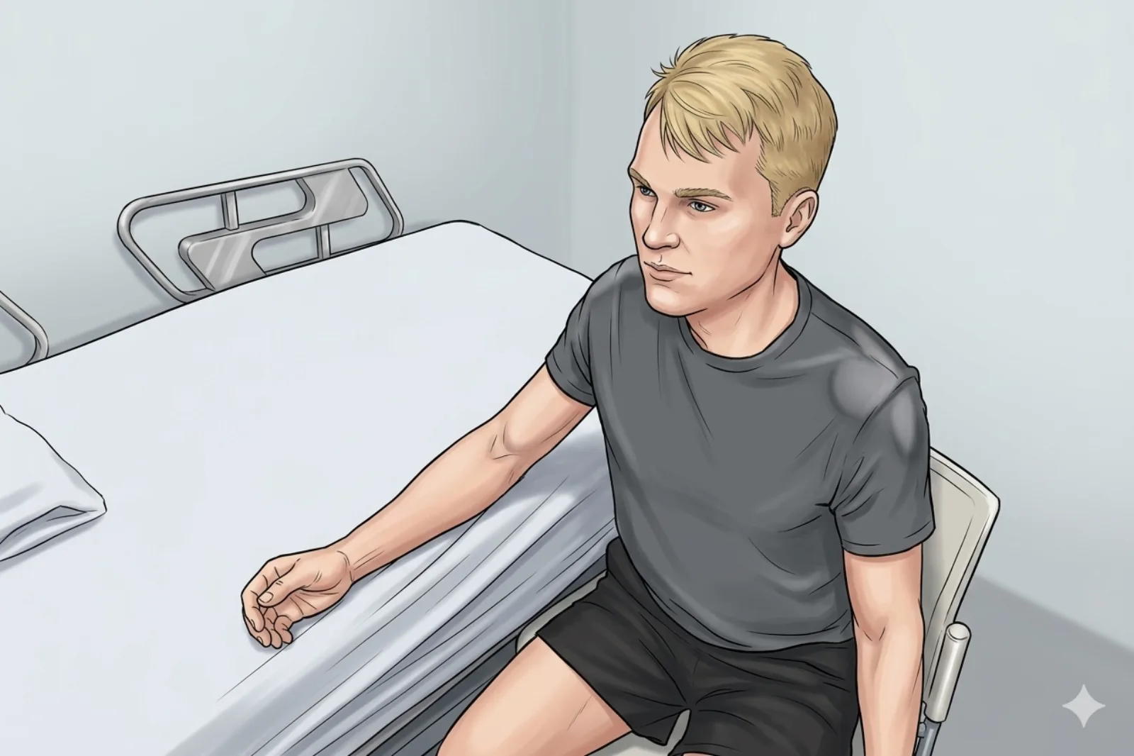
- Hand on table; thumb in relaxed/neutral position with radial aspect up; slight wrist ulnar deviation to open the anatomical snuffbox; small towel under wrist
Probe
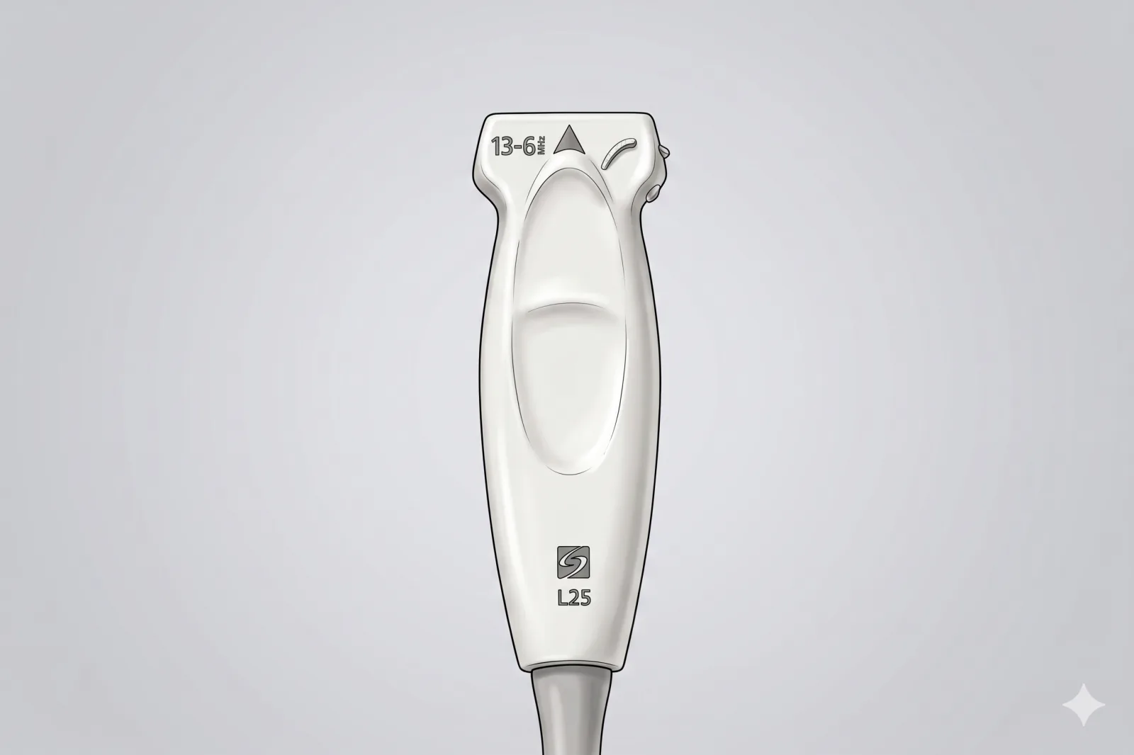
- High-frequency linear probe (15+ MHz hockey-stick if available)
Landmarking — find the view
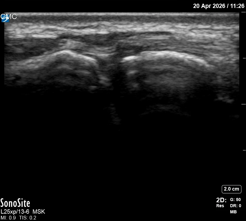
- Slide distal to the radial styloid and identify the trapezium → 1st metacarpal base joint, between the APL/EPB tendons (volar border) and the EPL tendon (dorsal border).
- Center the joint cleft and Doppler the radial artery (lies just deep/dorsal in the snuffbox).
The correct ultrasound view
- Two cortical bones (trapezium and 1st MC base) with thin anechoic joint line; APL/EPL on either side
Needle corridor
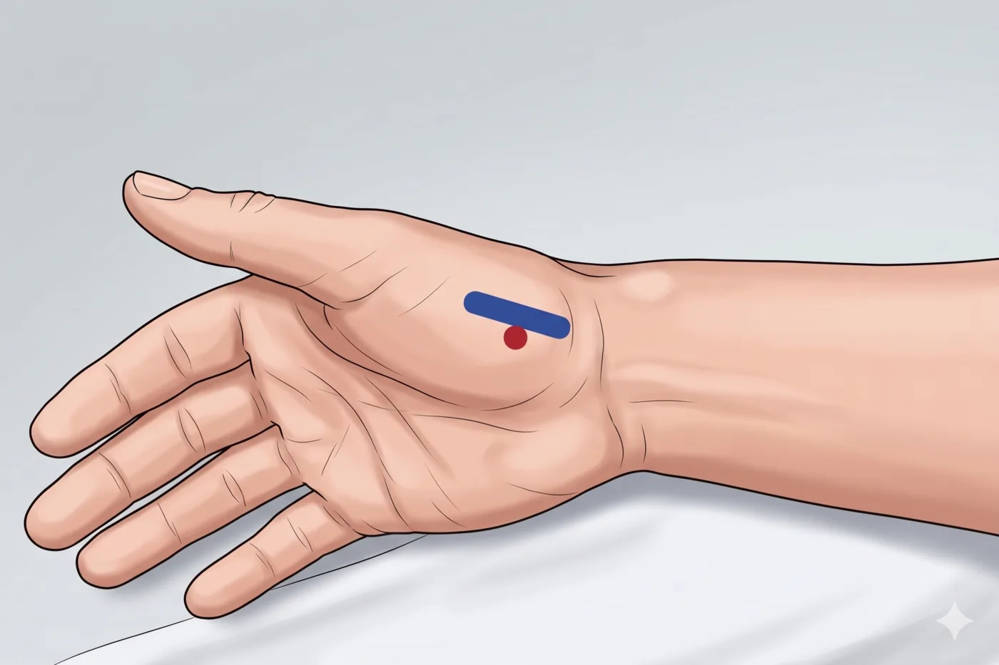
- Dorsal in-plane approach distal-to-proximal along the joint plane, between APL and EPL tendons
Avoid
- Radial artery (deep in the snuffbox — Doppler before entry)
- Dorsal sensory branches of the radial nerve
- Intratendinous APL or EPL placement
Steps
- 1Advance in-plane into the joint cleft; inject up to 1 mL slowly.
- 2STOP IMMEDIATELY if the patient reports electric, shooting, or paresthetic sensations into the thumb.
Tips
- Tiny joint — total injectate ≤ 1 mL. Don't waste space with too much local first.
- Limit to ≤ 3 injections per joint per year.
- If color Doppler shows the radial artery in the planned path, abandon and re-plan.
Resident pearls
- Identify the 1st CMC joint by sliding distal to the radial styloid; avoid the radial artery in the snuffbox.
- The target is the 'joint gap' — confirm flow with capsular distension.
- Limited volume — pre-mix steroid and a tiny lidocaine aliquot in the same syringe.
Supplies
- 3 mL Syringe
- 27G 0.5-1" Needle
- 18G Drawing Needle
- 1% Lidocaine (0.3-0.5 mL)
- Dexamethasone 4 mg (PREFERRED — non-particulate, non-particulate; lower risk of skin and fat atrophy at this superficial joint) OR triamcinolone 10-20 mg
- Alcohol Swabs (vial tops)
- Betadine/Chloraprep
- Sterile Gloves
- Sterile 4x4 Gauze Pads
- Sterile Towels & Drapes
- Adhesive Bandage (Band-Aid)
- Washcloth/Towel (gel cleanup)
- Needle Disposal Safety Cap
- Sterile probe cover & gel
Post-procedure
- Apply a clean dressing; remove after 24 hours.
- Local anesthetic relief for 1-4 hours; steroid begins 24-72 hours.
- Steroid 'flare' in 2–10% — manage with ice and acetaminophen.
- Rest the thumb 24-48 hours; thumb spica splint optional for comfort.
- Return immediately for: spreading redness, swelling, fever, disproportionate pain, OR thumb/finger color change or coolness (concern for vascular compromise).
- Diabetics: expect 50-100 mg/dL glucose rise for 3-5 days.
- Maximum 3 injections per year.