Ultrasound · Hip
Greater Trochanteric Pain Syndrome — Peritendinous Injection
hipgluteus mediustendinopathytrochanteric
Treats
- Greater trochanteric pain syndrome (gluteus medius/minimus tendinopathy ± peritendinous fluid). True isolated bursitis is uncommon.
Contraindications
- Active local skin infection at entry site (absolute)
- Suspected septic bursa (absolute)
- Known allergy to local anesthetic, steroid, or antiseptic prep
- Therapeutic anticoagulation — review case-by-case
- Uncontrolled diabetes (relative)
- High-grade gluteus medius/minimus tendon tear (relative — favor regenerative injectate over particulate steroid)
Pre-procedure checklist
- Confirm patient identity (two identifiers)
- Confirm procedure, side, and site — mark with patient awake
- Review allergies and anticoagulation
- Confirm informed consent
- Pre-scan: identify GMx, IT band, gluteus medius/minimus tendons; Doppler superior gluteal branches
- Time-out immediately before needle entry
Positioning
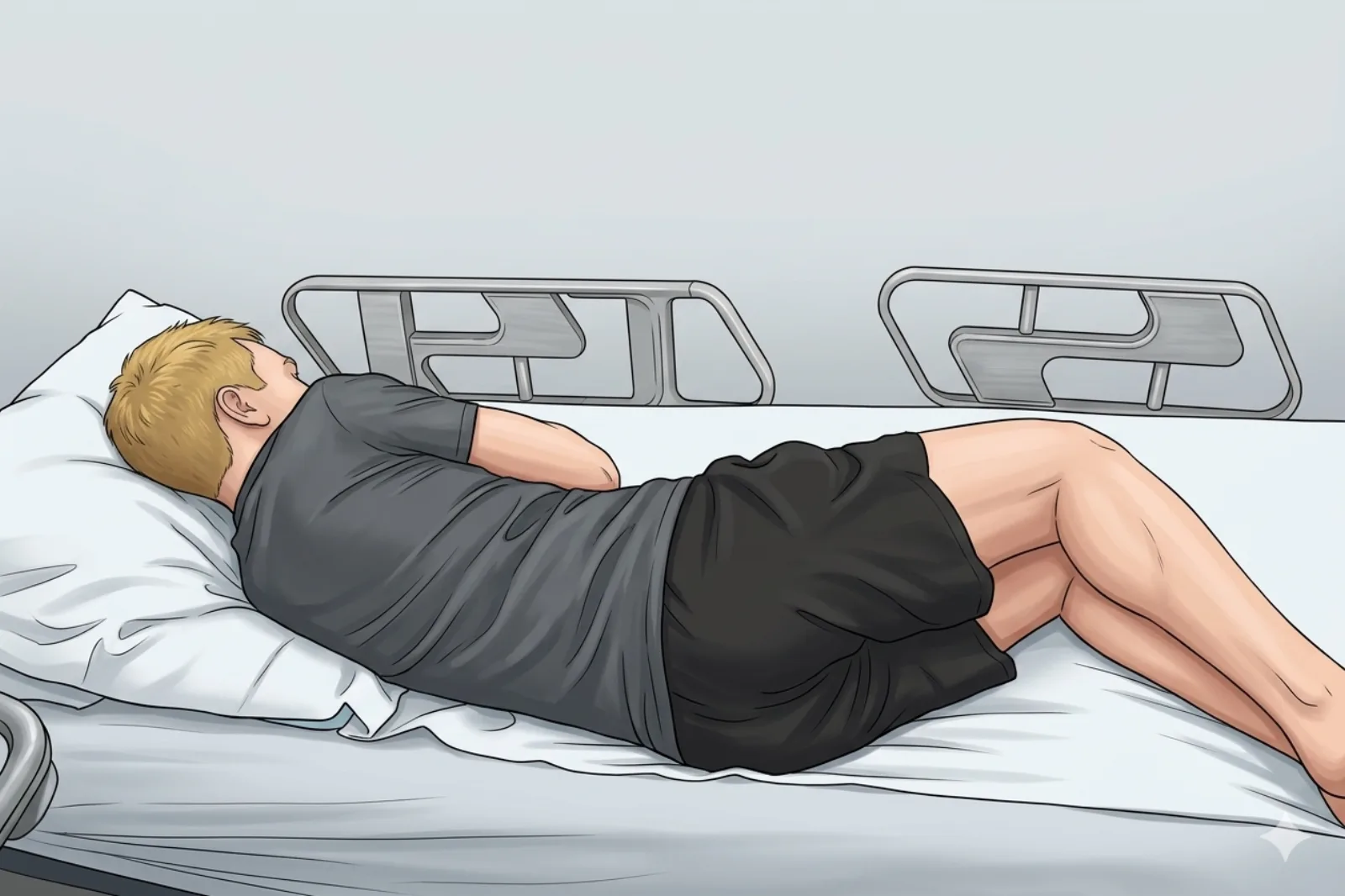
- Side-lying, symptomatic side up
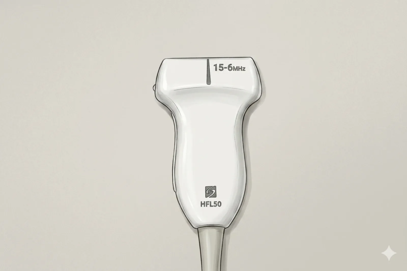
Probe
- Linear or curvilinear depending on habitus
Landmarking — find the view
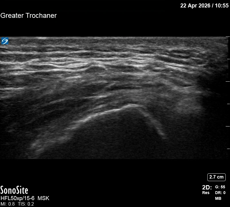
- Visual Cue: The 'Apex of the Hill' — Identify the bright convex prominence of the greater trochanter.
- Slide posteriorly to visualize the gluteus medius and minimus tendons.
- Identify the IT Band as the superficial 'roof' over the trochanteric hill.
- Find the hypoechoic plane (the bursa/peritendinous space) between the IT band and the gluteus medius/minimus tendons.
The correct ultrasound view
- Distinct dark plane separating superficial IT band/GMx from deep gluteus medius/minimus tendon
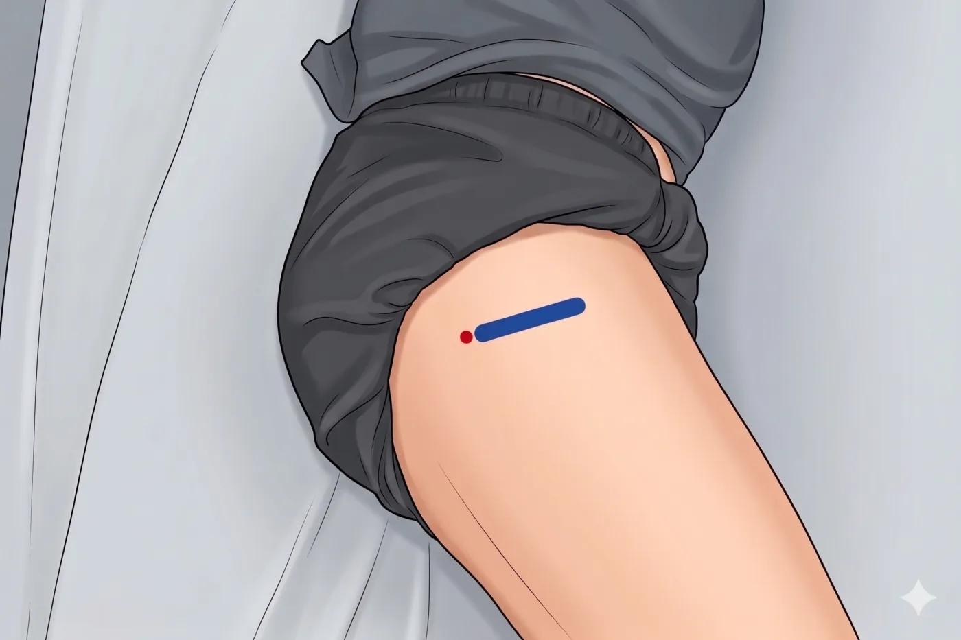
Needle corridor
- In-plane into the peritendinous plane: IT band/GMx (above), gluteus medius/minimus tendon (below)
Avoid
- Superior gluteal artery branches (ascending/transverse branches cross the peritrochanteric region variably) — Doppler before every entry. No large bundles cross directly over the trochanteric apex, but branches are variable.
- Sciatic nerve — runs posteriorly in the gluteal region; if the needle angles too deep or posteriorly during the lateral approach, the sciatic nerve may be approached. Keep the probe and needle trajectory lateral and superficial to the peritendinous plane.
Steps
- 1In-plane into peritendinous plane.
- 2Inject 3-5 mL into the peritendinous plane (tendinopathy target); up to 8 mL only for true bursal hydrodissection.
- 3Confirm smooth spread; ensure the IT band lifts off the tendon.
- 4STOP IMMEDIATELY if the patient reports electric, shooting, or paresthetic sensations into the leg.
Tips
- Slight rotation in line with GMx fibers may increase conspicuity.
- Limit corticosteroid to ≤ 3 per site per year.
- If color Doppler shows a vessel directly in the planned path, abandon and re-plan.
Resident pearls
- The peritendinous plane is the hypoechoic space between the IT band and the gluteus medius/minimus tendons.
- Rotate the probe to align with gluteus maximus fibers to see the plane more clearly.
- Ensure the IT band 'lifts off' the tendon during injection.
- Modern terminology: 'greater trochanteric pain syndrome' (gluteal tendinopathy ± peritendinous fluid) — true isolated bursitis is uncommon.
Supplies
- 10 mL Syringe
- 22G 1.5" or 3.5" Needle
- 18G Drawing Needle
- 1% Lidocaine (3-5 mL)
- Triamcinolone 40 mg OR Dexamethasone 4-10 mg
- Alcohol Swabs (vial tops)
- Betadine/Chloraprep
- Sterile Gloves
- Sterile 4x4 Gauze Pads
- Sterile Towels & Drapes
- Adhesive Bandage (Band-Aid)
- Washcloth/Towel (gel cleanup)
- Needle Disposal Safety Cap
- Sterile probe cover & gel
Post-procedure
- Apply a clean dressing; remove after 24 hours.
- Local anesthetic relief for 1-4 hours; steroid begins 24-72 hours.
- Steroid 'flare' in 2–10% — manage with ice and acetaminophen.
- Rest the hip 24-48 hours; resume normal activity as tolerated thereafter.
- Return immediately for: spreading redness, swelling, fever, disproportionate pain, or new leg weakness.
- Diabetics: expect 50-100 mg/dL glucose rise for 3-5 days.
- Maximum 3 injections per site per year.