Ultrasound · Hip
Piriformis Injection
hippiriformissciatic nervedeep gluteal pain
Treats
- Piriformis syndrome with sciatic irritation
- Deep gluteal pain / non-spinal sciatica
Contraindications
- Active local skin infection at entry site (absolute)
- Known allergy to local anesthetic, steroid, or antiseptic prep
- Therapeutic anticoagulation — review case-by-case (deep gluteal injection)
- Uncontrolled diabetes (relative)
- Beaton-Anson sciatic variant with sciatic nerve or its peroneal division passing through the piriformis belly (relative — abandon intramuscular injection, use peri-fascial only)
- Active sciatic neuropathy from another cause (relative — re-evaluate diagnosis)
Pre-procedure checklist
- Confirm patient identity (two identifiers)
- Confirm procedure, side, and site — mark with patient awake
- Review allergies and anticoagulation
- Confirm informed consent
- Pre-scan: identify piriformis (confirm sacral attachment to distinguish from gemelli/obturator internus); identify sciatic nerve completely; check for Beaton-Anson variant (~15-20%); Doppler inferior gluteal vessels
- Time-out immediately before needle entry
Positioning
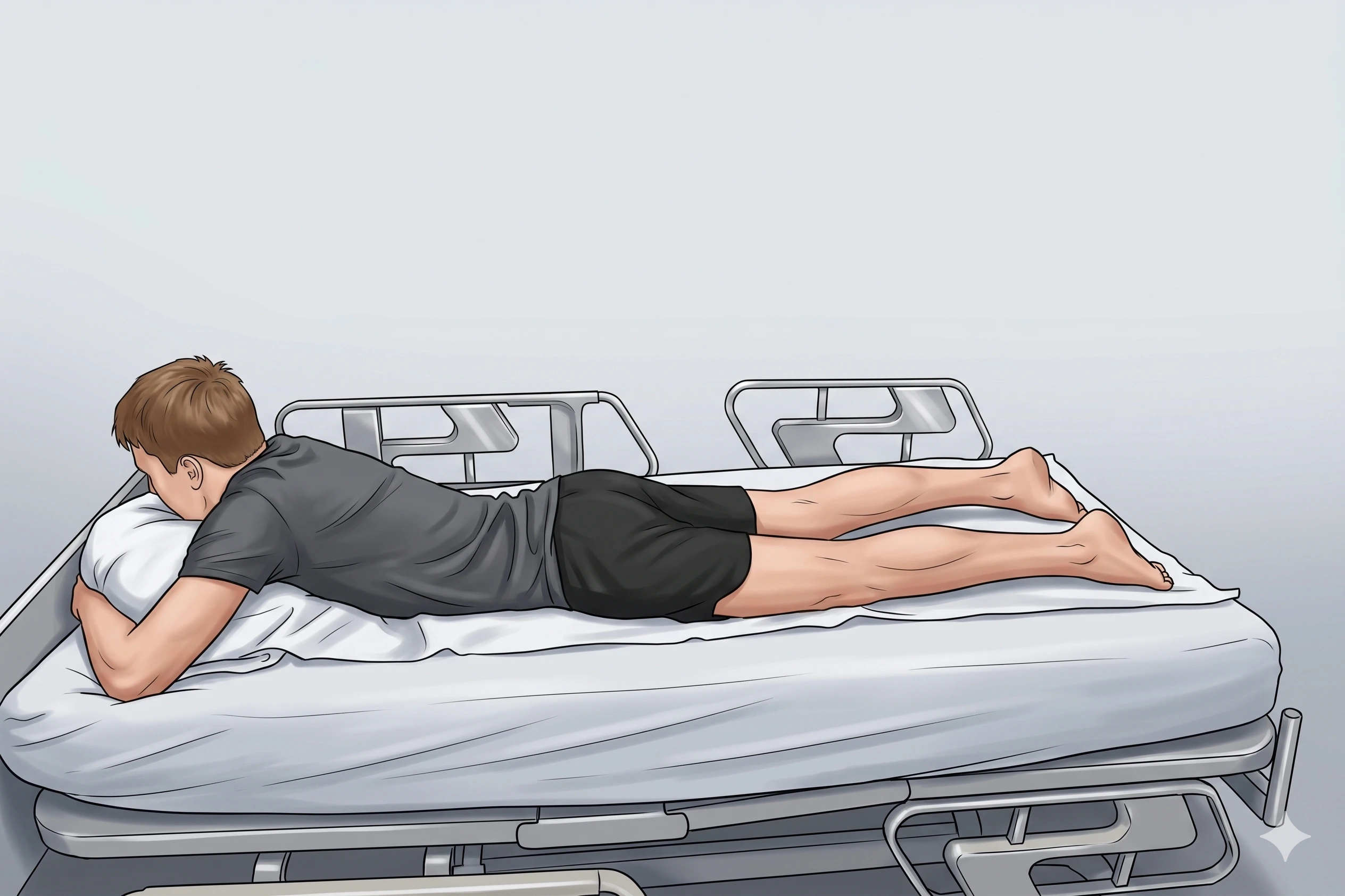
- Prone with neutral pelvis; pillow under iliac crests to improve visualization
Probe
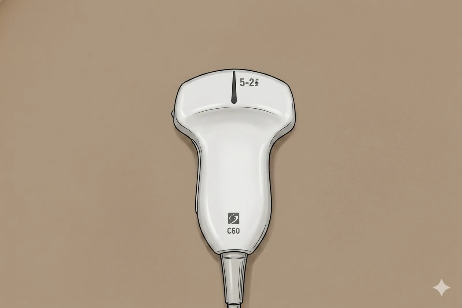
- Curvilinear probe (2-5 MHz) for depth; low-frequency linear in thin patients
Landmarking — find the view
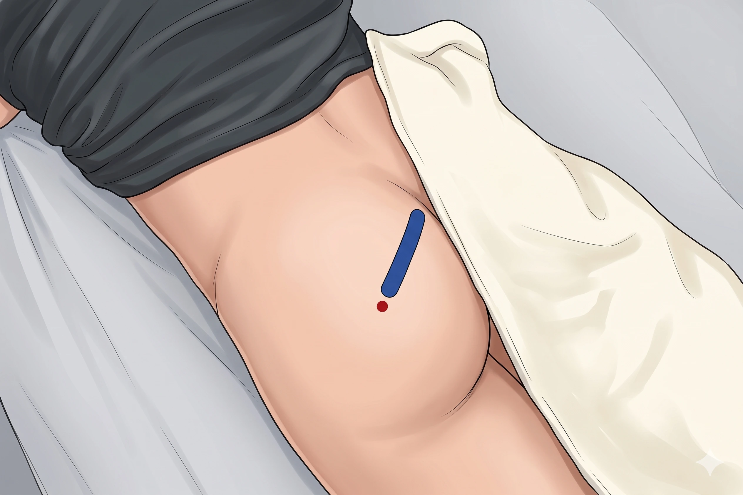
- Probe over PSIS → slide inferior to see sacrum medially and PIIS laterally
- Continue distally until ilium disappears (greater sciatic notch)
- Identify piriformis deep to gluteus maximus, coursing sacrum → greater trochanter
- Internally/externally rotate hip to confirm piriformis motion
- Identify sciatic nerve deep/inferior to piriformis along its full length before injection
- Confirm muscle is piriformis by tracing fibers MEDIALLY to the lateral sacrum (only piriformis attaches there — gemelli/obturator internus do not)
The correct ultrasound view
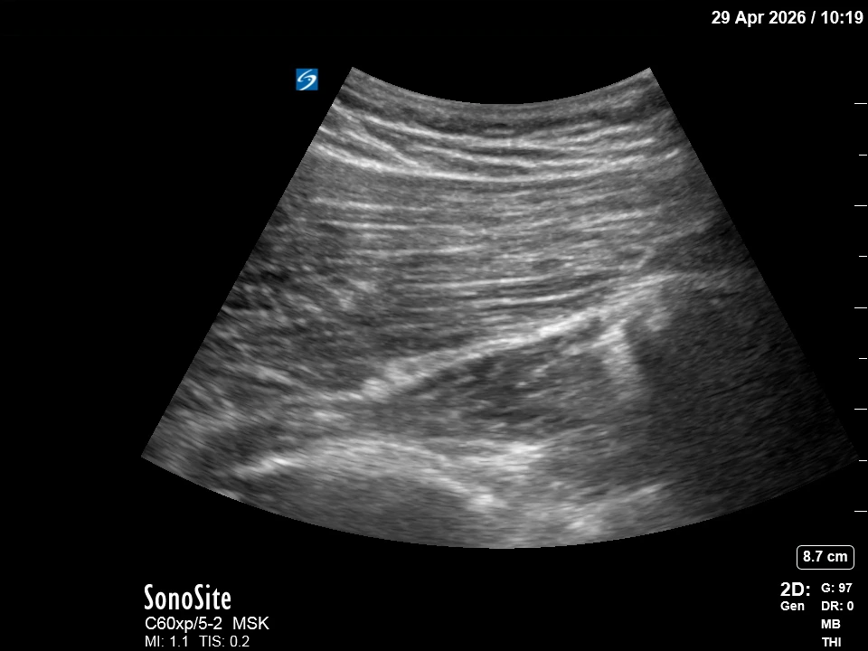
- Gluteus maximus superficial; fusiform piriformis belly oblique with clear sacral attachment; sciatic nerve deep (honeycomb)
Needle corridor
- Medial → lateral so the needle terminates lateral to the sciatic nerve and lateral to the inferior gluteal vessels. Between gluteus maximus (above) and piriformis (below); avoid sciatic nerve.
Avoid
- Sciatic nerve (always identify before injection — in Beaton-Anson variants the nerve passes through the muscle)
- Inferior gluteal artery/vein
- Inferior gluteal nerve (L5, S1, S2 — exits through greater sciatic foramen below piriformis and innervates gluteus maximus; injury causes gluteus maximus weakness)
- Posterior femoral cutaneous nerve (runs with inferior gluteal vessels)
- Pudendal NV bundle (if advancing medially toward ischial spine)
Steps
- 1Advance in-plane (medial → lateral) under real-time US.
- 2Stop at piriformis belly or perifascial plane lateral to the sciatic nerve; aspirate gently.
- 3Test inject a small bolus; then inject anesthetic ± steroid or botulinum toxin as indicated.
- 4STOP IMMEDIATELY if the patient reports electric, shooting, or paresthetic sensations down the leg — this indicates intraneural or perineural sciatic placement. Withdraw 1-2 mm and reposition.
Tips
- Map vessels with Doppler; if sciatic nerve not clearly visualized along its full course, do not inject.
- If color Doppler shows a vessel in the planned path, abandon and re-plan.
- Limit corticosteroid to ≤ 3 per site per year.
Resident pearls
- Beaton-Anson variants: In ~15-20% of patients the sciatic nerve or its peroneal division courses THROUGH the piriformis belly. Before any intramuscular injection, scan the entire piriformis and trace the sciatic nerve continuously — if the nerve passes through the muscle, abandon intramuscular injection and use a peri-fascial approach only.
- The most common identification error is confusing piriformis with the superior/inferior gemellus or obturator internus immediately caudal. All four look similar. The piriformis is the ONLY one whose fibers clearly attach to the lateral sacrum — always trace medially to confirm.
- Confirm the piriformis muscle by having the patient internally/externally rotate the hip.
- Approach medial-to-lateral so the needle terminates lateral to the sciatic and lateral to the inferior gluteal vessels.
Supplies
- 10 mL Syringe
- 22G 3.5" Spinal Needle
- 18G Drawing Needle
- 1% Lidocaine (5-10 mL)
- Triamcinolone 40 mg OR Dexamethasone 4-10 mg
- If using botulinum toxin: onabotulinumtoxin A 100 U typical (range 50-200 U). Dosing is product-specific and NOT interchangeable between brands (Botox ≠ Dysport ≠ Xeomin) — confirm product, dilution, and dose with attending before drawing up.
- Alcohol Swabs (vial tops)
- Betadine/Chloraprep
- Sterile Gloves
- Sterile 4x4 Gauze Pads
- Sterile Towels & Drapes
- Adhesive Bandage (Band-Aid)
- Washcloth/Towel (gel cleanup)
- Needle Disposal Safety Cap
- Sterile probe cover & gel
Post-procedure
- Apply a clean dressing; remove after 24 hours.
- Local anesthetic relief for 1-4 hours; steroid begins 24-72 hours.
- Steroid 'flare' in 2–10% — manage with ice and acetaminophen.
- Rest 24-48 hours; resume normal activity as tolerated thereafter.
- Return immediately for: spreading redness, swelling, fever, disproportionate pain, OR new leg weakness, foot drop, or worsening sciatica.
- Diabetics: expect 50-100 mg/dL glucose rise for 3-5 days.
- Maximum 3 injections per site per year.