Ultrasound · Knee
Iliotibial Band (IT Band) Peritendinous Injection
kneeIT bandiliotibial bandITBSlateral kneelateral femoral epicondyleLFErunner's kneeperitendinousfriction syndromeGerdy tubercledistal ITB
Treats
- Iliotibial band syndrome (ITBS) — lateral knee pain from inflammation of innervated loose connective/fat tissue deep to the distal ITB at the lateral femoral epicondyle (impingement zone at 20–30° flexion)
- Distal ITB tendinopathy or insertional enthesopathy at Gerdy tubercle (peritendinous approach)
- Diagnostic anesthetic block to confirm LFE as pain generator vs lateral meniscus, popliteus, LCL, or biceps femoris pathology
- Snapping IT band over the LFE with associated peritendinous fluid
Contraindications
- Active cellulitis or skin infection over the lateral knee entry site (absolute)
- Known allergy to planned local anesthetic, corticosteroid, or antiseptic prep (absolute)
- Bacteremia or systemic sepsis (absolute)
- Septic bursitis or septic joint — aspirate and culture first; do not inject steroid (absolute)
- Therapeutic anticoagulation — 25G peritendinous injection is low-risk but individualize per ASRA/AIUM guidance (relative)
- Poorly controlled diabetes (HbA1c >8–9%) — counsel on transient glycemic rise (relative)
- Prior corticosteroid injection to the same site within 3 months — risk of fat atrophy and skin depigmentation is elevated given how superficial the ITB target is (relative)
Pre-procedure checklist
- Confirm patient identity with two identifiers (name and DOB)
- Confirm procedure, laterality, and mark the site with the patient awake
- Review allergies (local anesthetic, corticosteroid, latex, chlorhexidine/iodine) and anticoagulation status
- Confirm and document informed consent — warn specifically about fat atrophy and skin depigmentation given the superficial target
- Pre-scan the lateral knee over the LFE: identify the hyperechoic fibrillar ITB, hypoechoic peritendinous fat/fluid, and bony LFE contour
- Apply color/power Doppler to map the lateral superior and lateral inferior genicular arteries before planning the trajectory
- Time-out immediately before needle insertion (patient, site, laterality, procedure, medication, dose confirmed aloud)
Positioning
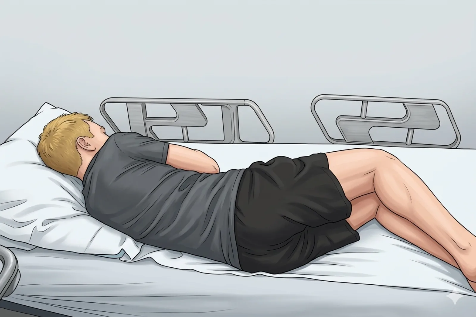
- Patient lying on side (lateral decubitus), affected side UP on the exam table.
- Contralateral hip and knee flexed ~45–60° for stability; place a pillow between the knees.
- Affected knee flexed to approximately 20–30° — the impingement angle where the ITB tensions over the LFE and peritendinous fluid is maximally accessible.
- Support the lower leg on a pillow so the tibia is parallel to the table; lateral aspect of the knee faces directly upward toward the operator.
Probe
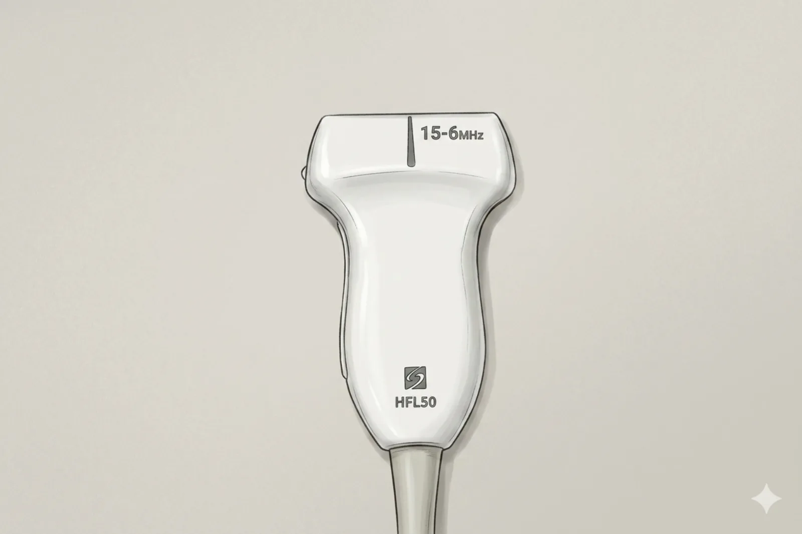
- High-frequency linear probe, 10–18 MHz (12–18 MHz hockey-stick small-footprint preferred for the superficial lateral LFE target)
- Depth 2.0–3.0 cm; focal zone at 5–10 mm (target lies within 3–8 mm of skin in most patients)
Landmarking — find the view
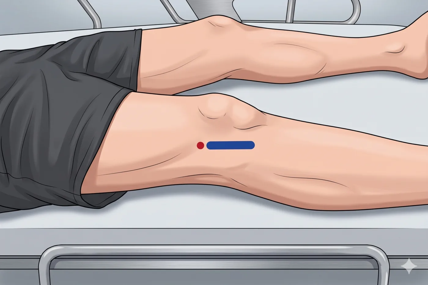
- Palpate the lateral femoral epicondyle (LFE) — the prominent bony ridge on the lateral distal femur, proximal to the lateral joint line.
- Place the probe in long axis (longitudinal) to the ITB fibers centered over the LFE; identify the thin hyperechoic fibrillar ITB running parallel to skin.
- Deep to the ITB, identify the hypoechoic loose connective tissue / fat (or discrete anechoic pseudo-bursa in active ITBS) between the ITB and the convex LFE cortex.
- Slide the probe proximal and distal to find the point of maximal peritendinous thickening or fluid — usually just proximal to or directly over the LFE apex.
- Rotate 90° to short axis to confirm the crescentic fluid collection between ITB and bone.
- Apply color Doppler (low PRF) to map the lateral superior genicular artery (proximal to LFE) and lateral inferior genicular artery (near joint line/fibular head) before planning the needle trajectory.
The correct ultrasound view
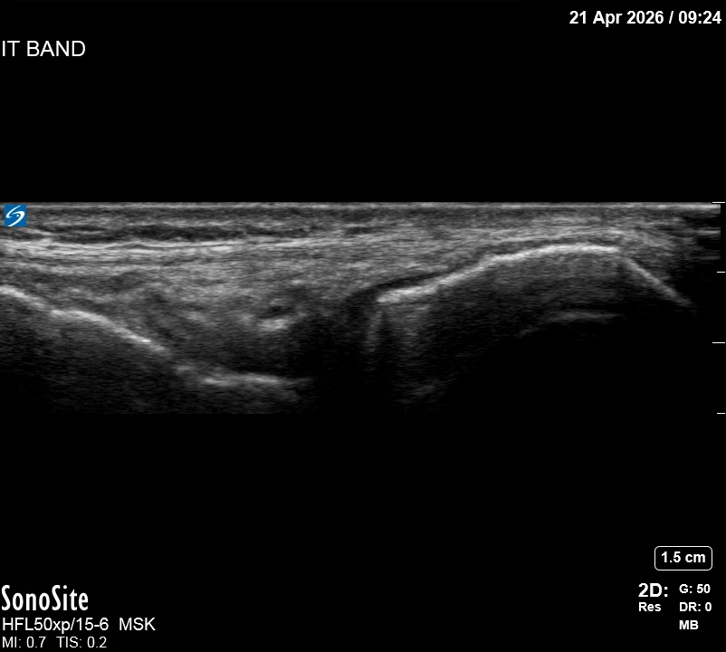
- Long axis: convex hyperechoic LFE cortex deep; immediately superficial, a hypoechoic layer of loose connective tissue or anechoic fluid (the injection target); superficial to that, the thin linear hyperechoic fibrillar ITB running parallel to skin.
- On dynamic scan through 20–40° of flexion (the impingement zone), the ITB should glide smoothly over the LFE; restricted glide or tethering supports the diagnosis.
- With color Doppler, the peritendinous plane should be avascular — any vessel crossing the planned trajectory requires re-planning.
Needle corridor
- Peritendinous plane between the deep surface of the ITB and the cortex of the lateral femoral epicondyle — specifically into the hypoechoic loose connective tissue / fat / pseudo-bursa.
- Injectate should hydrodissect (lift) the ITB off the LFE cortex on real-time imaging.
- Do NOT inject intratendinously into the ITB substance — this risks tendon weakening and misses the pain generator.
- Do NOT inject into subcutaneous tissue superficial to the ITB — risk of fat atrophy and skin depigmentation.
Avoid
- Lateral superior genicular artery — runs transversely across the distal-lateral femur just proximal to the LFE; always Doppler before entry
- Lateral inferior genicular artery — near the fibular head and lateral joint line; at risk with distal trajectories
- Common peroneal (fibular) nerve — wraps the fibular neck posterolaterally, 3–5 cm distal/posterior to the LFE; avoid posterior and distal trajectories
- Lateral collateral ligament (LCL) — runs from LFE to fibular head; do not traverse
- Biceps femoris tendon — inserts on fibular head posterior to LCL
- Subcutaneous fat superficial to the ITB — high risk of steroid-induced fat atrophy and skin hypopigmentation
Steps
- 1Position patient in lateral decubitus with affected knee flexed 20–30°.
- 2Pre-scan, map genicular vessels with color Doppler, mark skin entry, and complete time-out.
- 3Sterile prep and drape the lateral knee; apply sterile probe cover and gel.
- 4Place probe in long axis to the ITB over the LFE.
- 5Raise a small skin wheal of 1% lidocaine (0.5–1 mL) with a 27–30G needle at the planned entry point — inject only into skin, not into the peritendinous plane.
- 6Advance a 25G 1.5-inch needle in-plane along the ITB long axis at a shallow angle (20–40° to skin), entering from distal-to-proximal or proximal-to-distal based on vessel map.
- 7Advance the needle to the deep surface of the ITB without piercing its fibers — approach from the distal or proximal edge and slide the bevel into the hypoechoic peritendinous plane between the deep ITB surface and the LFE cortex.
- 8Inject a test bolus of 0.2–0.5 mL: the ITB should visibly lift off the bone with smooth anechoic spread. Tissue balling within the ITB = intratendinous; skin bleb = subcutaneous — reposition before continuing.
- 9Aspirate, then slowly inject the remaining 3–5 mL total over 20–30 seconds watching real-time spread.
- 10STOP IMMEDIATELY if patient reports shooting, electric, or paresthetic pain into the lower leg (common peroneal nerve proximity) or if resistance suddenly rises.
- 11Withdraw needle, apply pressure for 1–2 minutes, place adhesive bandage.
- 12Post-injection dynamic scan to confirm smooth ITB glide over the LFE through full range of motion.
Tips
- Shallow angle is critical — the target is 5–8 mm deep, so entry must be 1.5–2.5 cm away from the probe edge at 20–40° to the skin. A steep angle reduces needle visibility and risks plunging into cortex.
- Hydrodissect first, inject second — always confirm the plane opens with a 0.2–0.5 mL test bolus before committing the full volume.
- Doppler every time — lateral superior genicular artery anatomy is highly variable and frequently courses directly over the LFE.
- The ITB is one of the most superficial steroid targets at the knee — warn the patient specifically about fat atrophy and skin depigmentation; consider dexamethasone (non-particulate) if this is a concern.
- 25G is sufficient — the low-viscosity steroid/anesthetic mix does not require a larger bore; a smaller needle causes less post-procedure soreness in this superficial target.
Resident pearls
- "Lift the Band, Don't Stab the Band" — correct placement hydrodissects the ITB off the LFE. If the band doesn't lift, the tip is intratendinous.
- The 20–30° Rule — the ITB impinges on the LFE at this flexion angle (Orchard 1996, Fairclough 2006). Position the knee here to distend the peritendinous space.
- There is NO true bursa — Fairclough et al. (2006) showed the 'ITB bursa' is actually highly innervated loose connective/fat tissue, not a synovial bursa. The pain generator is this innervated fat layer.
- Injections treat symptoms, not mechanics — ITBS is fundamentally a hip-abductor weakness and overuse problem. Always refer to PT for gluteus medius strengthening and gait retraining, or symptoms recur in 6–12 weeks.
- Diagnostic anesthetic trick: inject 0.5–1 mL lidocaine alone first; resolution of pain on Noble compression test at 10 minutes confirms the ITB-LFE interface as the pain generator.
Supplies
- 25G 1.5" Needle (injection)
- 27-30G 0.5" Needle (skin wheal)
- 18G Drawing Needle
- 3 mL or 5 mL Syringe
- 1 mL Syringe (skin wheal)
- 1% Lidocaine (2–3 mL for injectate + 0.5–1 mL for skin wheal)
- Triamcinolone 20–40 mg OR Dexamethasone 4–8 mg (non-particulate preferred given superficial target)
- Alcohol Swabs (vial tops)
- Betadine/Chloraprep
- Sterile Gloves
- Sterile 4x4 Gauze Pads
- Sterile Towels & Drapes
- Adhesive Bandage (Band-Aid)
- Washcloth/Towel (gel cleanup)
- Needle Disposal Safety Cap
- Sterile probe cover & gel
- Pillow(s) for lateral decubitus positioning
Post-procedure
- Apply a clean adhesive bandage; keep it dry for 24 hours.
- Local anesthetic relief within 5–15 minutes, lasting 1–4 hours (lidocaine) or up to 6–8 hours (bupivacaine). Corticosteroid effect begins 24–72 hours after injection and peaks at 5–10 days.
- Steroid flare (2–10% of patients, typically 6–48 hours): manage with ice 15–20 minutes every 2–3 hours and acetaminophen or a short course of NSAIDs.
- Rest from impact activity (running, cycling, plyometrics) for 48–72 hours; gentle walking is fine immediately.
- Begin prescribed PT program: hip abductor/gluteus medius strengthening, single-leg stance, foam rolling the lateral thigh (avoid rolling directly over the LFE), and running-gait/cadence retraining.
- Diabetic patients: expect a 50–150 mg/dL rise in blood glucose for 3–7 days; monitor more frequently.
- Watch for and return immediately for: spreading redness, warmth, fever >38°C, purulent drainage, or disproportionate pain not controlled with ice and OTC analgesics.
- Warn the patient about possible skin depigmentation or a 1–2 cm area of subcutaneous fat atrophy appearing 1–3 months post-injection; usually cosmetic only and partially reverses over 6–12 months.
- Maximum of 3 corticosteroid injections per year at the same site, separated by at least 3 months.
- Follow-up at 4–6 weeks to reassess, confirm PT adherence, and plan next steps.