Ultrasound · Knee
Pes Anserine Bursa Injection (Medial Knee)
kneepes anserinemedial knee painbursitissartoriusgracilissemitendinosusSGTproximal medial tibiaMCLsaphenous nervetendinopathy
Treats
- Pes anserine bursitis — inflammation of the anserine bursa deep to the conjoined sartorius, gracilis, and semitendinosus (SGT) tendons at the proximal medial tibia
- Pes anserine tendinopathy or insertional enthesopathy of the conjoined SGT tendons
- Medial knee pain superimposed on osteoarthritis — pes anserine pain is present in 20–75% of patients with medial compartment OA
- Post-traumatic or post-surgical pes anserine pain, including after hamstring harvest for ACL reconstruction
- Diagnostic blockade to confirm the pes anserine as the pain generator before escalating management
Contraindications
- Overlying cellulitis, abscess, or skin breakdown at the planned needle entry site (absolute)
- Known or suspected septic bursitis or septic arthritis of the adjacent knee — aspirate first and culture (absolute)
- Documented allergy to planned local anesthetic, corticosteroid, or skin prep (absolute)
- Bacteremia or active systemic infection (absolute)
- Anticoagulation with supratherapeutic INR (>3.0) or recent therapeutic-dose DOAC (relative)
- Poorly controlled diabetes (HbA1c >8–9%) — corticosteroid will transiently raise glucose; counsel the patient (relative)
- Prior corticosteroid injection to the same site within 3 months (relative)
Pre-procedure checklist
- Confirm patient identity (two identifiers) and verify the diagnosis — tenderness 5–7 cm below the medial joint line at the pes insertion, pain with resisted internal rotation of the flexed knee
- Confirm procedure, laterality, and mark the site with the patient awake
- Review allergies (lidocaine, chlorhexidine/iodine, latex, corticosteroid preservatives) and anticoagulation status
- Confirm and document informed consent (infection, bleeding, post-injection flare, fat atrophy, skin hypopigmentation, tendon weakening, glycemic elevation in diabetics, vasovagal)
- Verify supplies and draw up medications into a labeled syringe; select needle length based on habitus (typically 22–25G 1.5 inch)
- Pre-injection diagnostic scan (non-sterile): identify the conjoined SGT tendons, pes anserine bursa (if distended), superficial MCL, and map the saphenous vein and inferior medial genicular vessels with color Doppler. Plan needle trajectory before sterile prep.
- Time-out immediately before needle insertion
Positioning
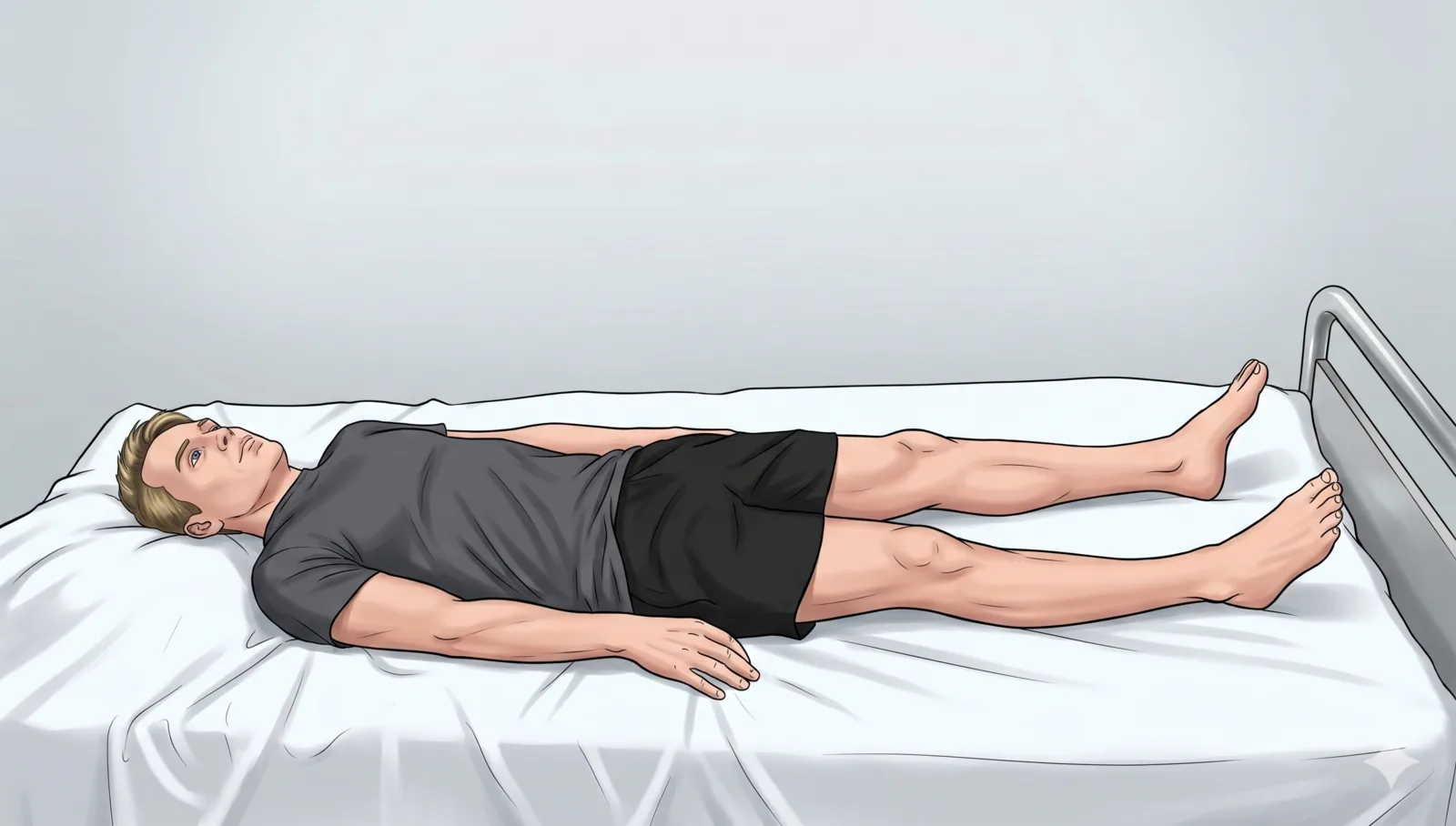
- Supine; leg extended; slight hip external rotation to expose the medial knee
- Operator stands at the medial side of the target knee; ultrasound machine on the contralateral side with screen at eye level
Probe
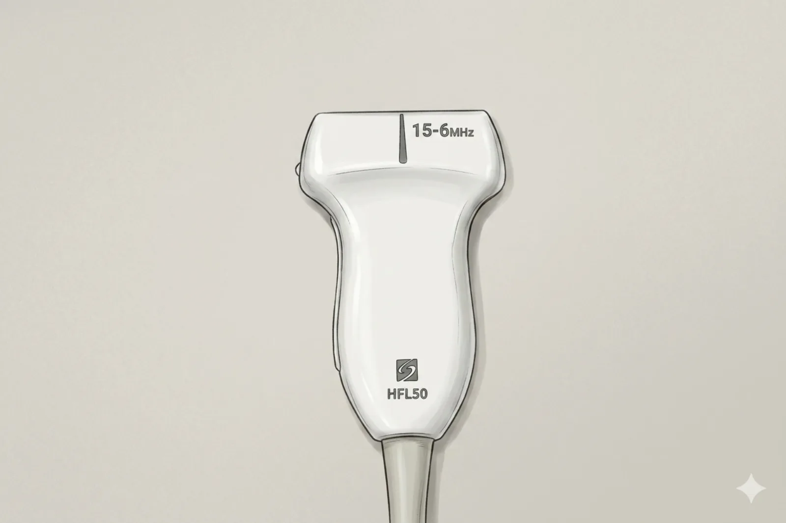
- High-frequency linear probe, 10–18 MHz (12–15 MHz standard linear or small-footprint hockey-stick preferred)
- Depth 2–4 cm; focal zone at 1–1.5 cm; MSK-superficial preset with compound imaging and needle enhancement enabled
Landmarking — find the view
- Find the medial joint line: probe in coronal long-axis over the medial knee, identify medial femoral condyle, medial meniscus, and medial tibial plateau.
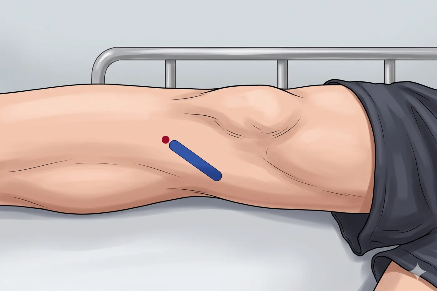
- Slide distally along the proximal medial tibia approximately 5–7 cm below the joint line in the coronal plane.
- Identify the superficial MCL — a thin crisp hyperechoic bilaminar band; this is the reference floor. The pes bursa lies superficial to the MCL.
- Rotate to short axis of the SGT tendons: three tendons appear as a single oval hyperechoic structure (sartorius most anterior/superficial, gracilis middle, semitendinosus most posterior/deep).
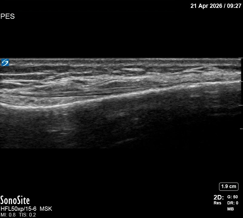
- Rotate 90° into long axis of the conjoined SGT tendon: fibrillar hyperechoic band coursing obliquely from posteromedial thigh to insert on the anteromedial proximal tibia ~5–7 cm below joint line.
- Identify the pes anserine bursa (if pathologic): anechoic or hypoechoic fluid collection between the deep SGT surface and the MCL/tibial periosteum. Normal bursae are often not visible.
- Apply color Doppler to locate the saphenous vein (anterior and superficial to the pes) and inferior medial genicular artery. Plan the entry from the posterior side to keep the trajectory away from the saphenous neurovascular bundle.
The correct ultrasound view
- Long axis of SGT tendon: from superficial to deep — skin/fat, conjoined SGT tendon (fibrillar hyperechoic band coursing obliquely to tibial insertion), pes anserine bursa (anechoic crescentic space if distended), superficial MCL (crisp hyperechoic bilaminar band), proximal medial tibial cortex (bright hyperechoic line with posterior shadowing).
- Short axis of SGT: three tendons as a flattened hyperechoic oval lying on top of the MCL, with the bursa as a thin hypoechoic half-moon deep to the tendon ensemble.
- Color Doppler should show no pulsatile vessel crossing the planned needle path.
Needle corridor
- Potential space between the deep surface of the conjoined SGT tendon and the superficial surface of the MCL/tibial periosteum at the pes insertion (~5–7 cm distal to the medial joint line).
- Needle enters from the posteromedial skin in-plane, directing distally and anteriorly along the deep surface of the tendon. This posterior-to-anterior trajectory keeps the needle away from the saphenous vein and nerve anteriorly.
- Do NOT inject into the SGT tendon substance (intratendinous = rupture risk and ineffective), deep to the MCL (joint entry), or into the saphenous vein/nerve territory.
Avoid
- Saphenous vein — superficial and anterior to the pes insertion; compressible on US, map with color Doppler
- Saphenous nerve — sartorial branch travels with the saphenous vein anteriorly; infrapatellar branch crosses the medial knee at the joint line. Both are at risk from anterior trajectories — use posteromedial approach.
- Superficial MCL — the 'floor' of the bursa; do not pierce it. If the needle dimples MCL fibers, withdraw 1–2 mm.
- Medial meniscus and medial joint capsule — more proximal and deeper; avoid advancing too proximally
- Inferior medial genicular artery and vein — near the joint line; visible with Doppler in thin patients
- SGT tendon substance — inject peritendinously or into the bursa, never intratendinously
- Tibial cortex / periosteum — withdraw 1–2 mm if needle tip contacts bone before injecting
Steps
- 1Sterile prep and drape: wide prep with 2% chlorhexidine/70% isopropyl alcohol; apply sterile probe cover and gel.
- 2Re-identify the target under the sterile probe in long-axis view of the SGT tendon; confirm the bursa, MCL, and absence of vessels in the planned path.
- 3Anesthetize: 25–27G needle with 1–2 mL of 1% lidocaine raises a skin wheal at the posteromedial entry site; infiltrate subcutaneously along the planned track without depositing into the bursa.
- 4Swap to the procedure needle: 22–25G 1.5-inch needle on a 3–5 mL syringe.
- 5Advance in-plane from the posteromedial aspect at a shallow angle (20–30° to skin); track the needle deep to the SGT tendon and superficial to the MCL, directing distally and anteriorly toward the pes insertion.
- 6Watch the needle tip at all times — if the shaft disappears, hold still and slide/tilt the probe to reacquire it before advancing further.
- 7Confirm bursal placement with hydrodissection: inject 0.5–1 mL anesthetic as a test bolus. Fluid should spread as a thin anechoic crescent between the tendon and MCL. If fluid disappears into the tendon (intratendinous) or spreads superficially (subcutaneous), reposition 1–2 mm and retest.
- 8Inject the therapeutic mixture: aspirate first, then inject slowly over 15–30 seconds (3–5 mL total). Injection should be low-resistance; high resistance means incorrect position — stop and reposition.
- 9STOP IMMEDIATELY if patient reports electric, shooting, or paresthetic pain into the lower leg (saphenous nerve proximity).
- 10Withdraw slowly while scanning for hematoma along the track; apply sterile bandage and firm local compression for 1–2 minutes.
- 11Immediate reassessment: have the patient perform the reproducing movement (seated-to-standing, stair step). >50% pain reduction within 10 minutes confirms the pes anserine as the pain generator.
Tips
- Enter posterior-to-anterior — the saphenous neurovascular bundle is anterior to the pes; entering from behind keeps the trajectory safely away from nerve and vein.
- Use hydrodissection as your tip-confirmation step, not anatomical confidence — a 0.5 mL test bolus of anesthetic shows you exactly where the tip is before committing the full dose.
- Defeat anisotropy before diagnosing pathology — the oblique SGT tendon course means small heel-toe probe tilts dramatically change tendon echogenicity. A 'hypoechoic' tendon that brightens with a 5° tilt is normal; one that stays dark is tendinopathic.
- Use color Doppler twice: once at the pre-scan to map the saphenous vein and genicular vessels, and again after injection to confirm no new hematoma.
- If no bursa is visible, inject peritendinously along the deep and superficial surfaces of the SGT tendon at the insertion — many patients with pes anserine syndrome have tendinopathy without a visible fluid collection.
Resident pearls
- "Say Grace before Tea" — Sartorius (anterior/superficial), Gracilis (middle), semiTendinosus (posterior/deepest). The bursa lies deep to all three at their common tibial insertion.
- Pes pain is 5–7 cm BELOW the medial joint line — lower than most residents expect. Medial joint line tenderness = meniscus or MCL, not pes. Palpating 4 fingerbreadths below the joint line along the anteromedial tibia reliably identifies the pes.
- 'Knees touching on the mattress' at night is a classic pes anserine history — the patient reports nocturnal medial knee pain when the painful knee presses into the bed or the other knee. A useful screening question.
- The bursa is often NOT visible on US even in symptomatic patients — a normal-appearing pes on ultrasound does not exclude bursitis. Correlate with the clinical exam and reproduction of the patient's pain.
- The superficial MCL is your floor — if your needle is above the MCL and the bursal space distends with your test bolus, you are in the correct plane. MCL fibers deforming = too deep, pull back 1–2 mm.
Supplies
- 22-25G 1.5" Needle (injection; 22G if aspirating thick fluid)
- 25-27G 1-1.5" Needle (skin wheal)
- 18G Drawing Needle
- 3-5 mL Luer-lock Syringe
- 3 mL Syringe (skin wheal)
- 1% Lidocaine (2–4 mL for injectate + 1–2 mL for skin wheal/subcutaneous track)
- Triamcinolone 20–40 mg OR Dexamethasone 4–10 mg (non-particulate preferred if skin pigmentation is a concern)
- Alcohol Swabs (vial tops)
- Betadine/Chloraprep
- Sterile Gloves
- Sterile 4x4 Gauze Pads
- Sterile Towels & Drapes
- Adhesive Bandage (Band-Aid)
- Washcloth/Towel (gel cleanup)
- Needle Disposal Safety Cap
- Sterile probe cover & gel
Post-procedure
- Observe 10–15 minutes post-injection for vasovagal reaction, allergic response, or immediate hematoma.
- Apply ice over the site 15–20 minutes, 2–3 times in the first 24 hours to reduce local soreness.
- Local anesthetic relief within 1–4 hours; pain may transiently return as anesthetic wears off before corticosteroid effect begins at 24–72 hours. Maximal benefit at 1–2 weeks.
- Steroid flare in 2–10% of patients — increased pain for 24–48 hours, peaking at 12–24 hours. Manage with ice and acetaminophen. Flare is NOT an infection.
- Return immediately for: redness spreading beyond the entry site, fever >38°C, increasing pain after 48 hours, purulent drainage, or systemic symptoms.
- Relative rest for 48 hours: avoid strenuous lower-extremity activity, running, deep squats, and stair climbing. Normal walking and daily activities are encouraged.
- Diabetic patients: monitor blood glucose 4 times daily for 5–7 days. Triamcinolone/methylprednisolone can raise glucose by 50–150 mg/dL for up to a week.
- Begin targeted rehabilitation at day 3–5: hamstring and adductor stretching, eccentric medial hamstring strengthening, gluteus medius activation, and correction of contributing biomechanics (valgus alignment, overpronation, weak hip abductors).
- Limit corticosteroid injections at this site to no more than 3 per year, separated by at least 3 months.
- Follow-up at 4–6 weeks. If <50% relief and exam still localizes to the pes, repeat ultrasound for occult pathology or reconsider alternate diagnosis.