Ultrasound · Knee
Suprapatellar Recess Injection
kneesuprapatellarrecessfat padprefemoralquadriceps
Treats
- Knee osteoarthritis
- Synovitis/effusion
- Diagnostic anesthetic
Contraindications
- Active local skin infection at entry site (absolute)
- Suspected septic joint (absolute — aspirate first, do not inject steroid)
- Bacteremia (absolute)
- Known allergy to local anesthetic, steroid, or antiseptic prep
- Therapeutic anticoagulation — review case-by-case
- Uncontrolled diabetes (relative)
- Knee prosthesis at the target site (relative — coordinate with orthopedics)
Pre-procedure checklist
- Confirm patient identity (two identifiers)
- Confirm procedure, side, and site — mark with patient awake
- Review allergies and anticoagulation
- Confirm informed consent
- Pre-scan: identify quadriceps fat pad, prefemoral fat pad, and the recess between them; Doppler genicular vessels
- Time-out immediately before needle entry
Positioning
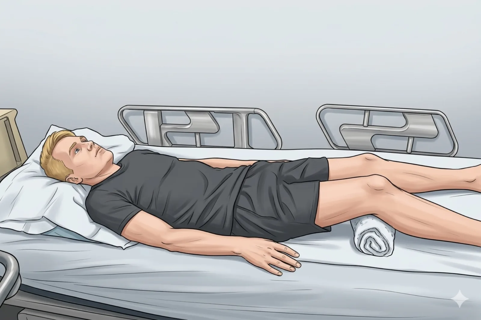
- Supine; small towel under knee for 20-30° flexion
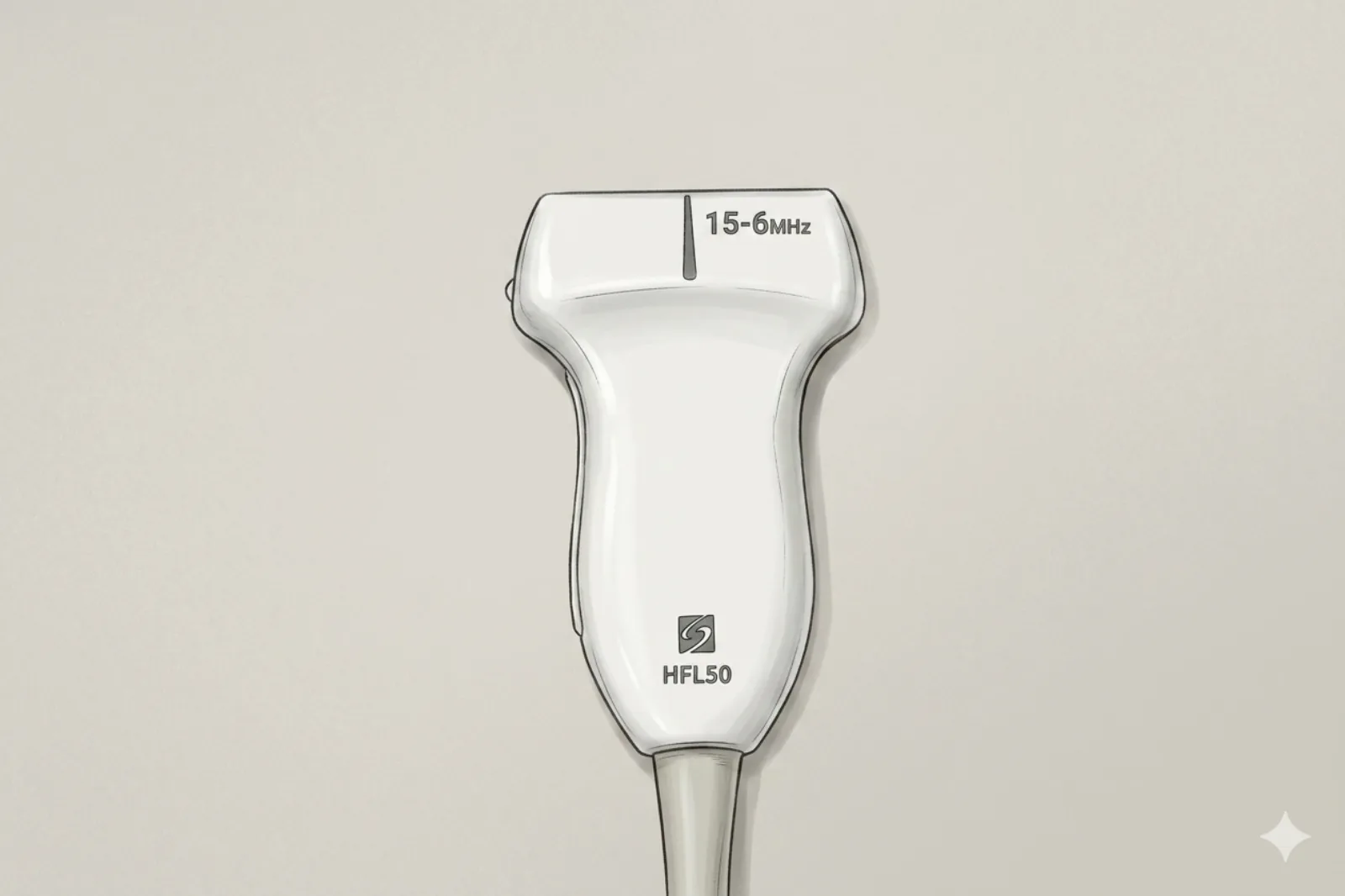
Probe
- High-frequency linear probe (6-13 MHz)
Landmarking — find the view
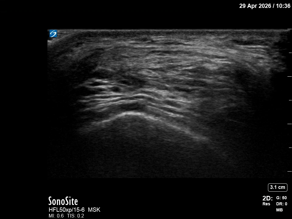
- Short-axis (transverse) probe across the suprapatellar recess just proximal to the patella → identify femoral cortex deep, prefemoral fat pad above, suprapatellar recess (target) above that, quadriceps fat pad and quadriceps tendon most superficial.
- Slide the probe to find the deepest part of the recess for easier access.
The correct ultrasound view
- Two distinct fat pads with anechoic space between — quadriceps fat pad (above) and prefemoral fat pad (below).
Needle corridor
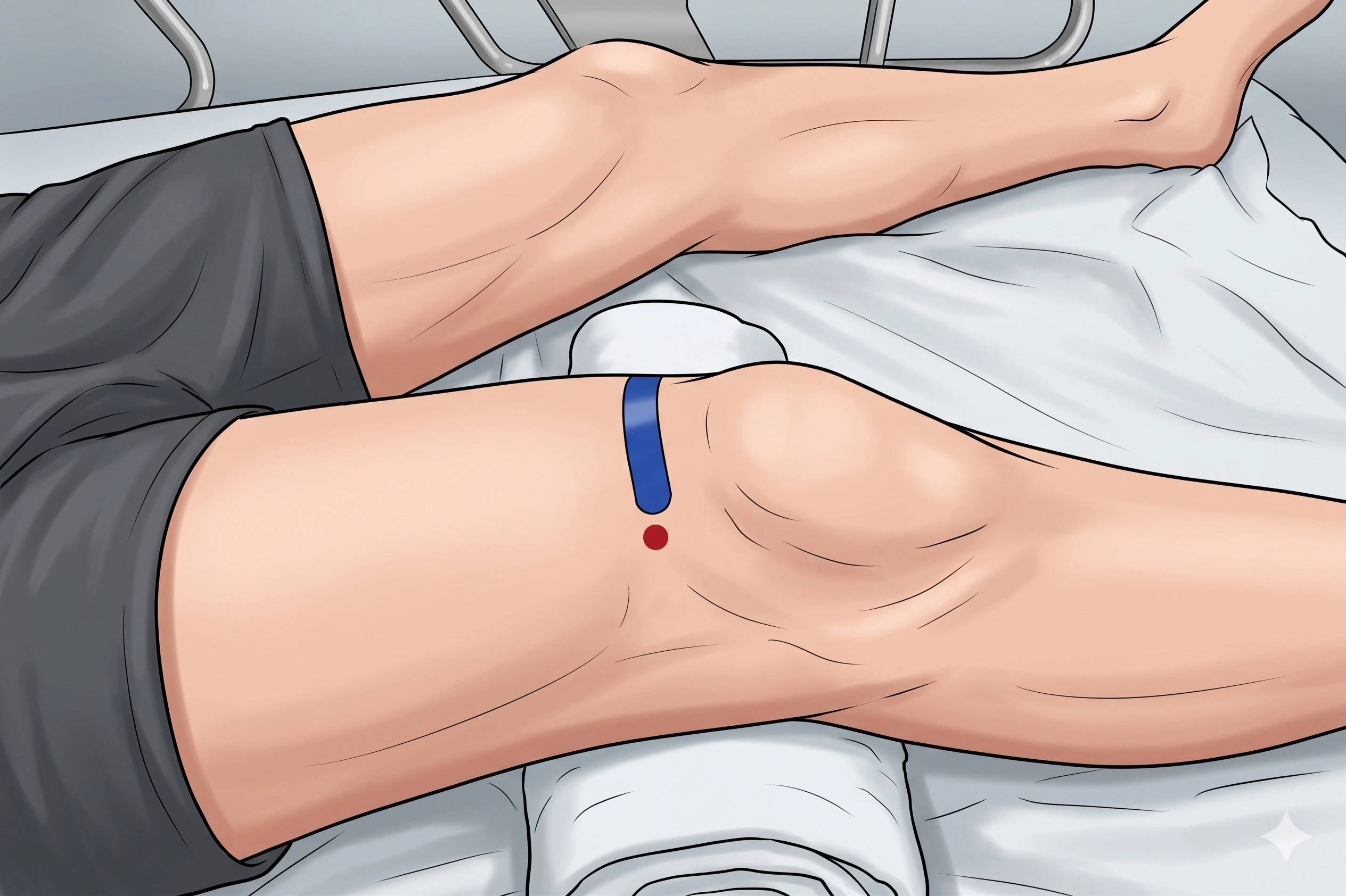
- Lateral → medial in-plane approach across the recess (with probe in transverse / short-axis orientation)
Avoid
- Avoid traversing the quadriceps tendon
- Genicular vessels (screen with Doppler)
Steps
- 1Position patient supine with knee in 20-30° flexion.
- 2Place probe in SHORT-AXIS (transverse) orientation across the suprapatellar recess just proximal to the patella.
- 3Identify the anechoic effusion or the hypoechoic plane between fat pads.
- 4Advance in-plane LATERAL → MEDIAL across the recess.
- 5Confirm spread with a small test bolus of local anesthetic.
- 6STOP IMMEDIATELY if the patient reports electric, shooting, or paresthetic sensations.
Tips
- Gentle probe compression pools fluid at your entry site; keep needle parallel to cartilage.
- Limit corticosteroid to ≤ 3 per joint per year.
- If color Doppler shows a genicular vessel directly in the planned path, abandon and re-plan.
Resident pearls
- The 'Two-Pad Sandwich': The target is the 'filling' between the Quadriceps Fat Pad (the top bun) and Prefemoral Fat Pad (the bottom bun).
- 20-30° Flexion: This 'pools' fluid into the suprapatellar space and relaxes the quad tendon.
- Short-axis transverse probe + lateral-to-medial in-plane = the standard approach. (A long-axis sagittal probe would require a superior-to-inferior in-plane entry instead — choose one, do not mix.)
- Gentle pressure with the probe can 'milk' fluid towards your needle tip.
Supplies
- For aspiration + injection: 18G or 20G 1.5" needle (large bore for viscous synovial fluid)
- For injection only (no aspiration): 25G 1.5" needle
- 10 mL or 20 mL Syringe
- 18G Drawing Needle
- 1% Lidocaine (5-10 mL)
- Triamcinolone 40 mg OR Dexamethasone 4-10 mg
- Alcohol Swabs (vial tops)
- Betadine/Chloraprep
- Sterile Gloves
- Sterile 4x4 Gauze Pads
- Sterile Towels & Drapes
- Adhesive Bandage (Band-Aid)
- Washcloth/Towel (gel cleanup)
- Needle Disposal Safety Cap
- Sterile probe cover & gel
Post-procedure
- Apply a clean dressing; remove after 24 hours.
- Local anesthetic relief for 1-4 hours; steroid begins 24-72 hours.
- Steroid 'flare' in 2–10% — manage with ice and acetaminophen.
- Rest the knee 24-48 hours; resume normal activity as tolerated thereafter.
- Return immediately for: spreading redness, swelling, fever, or disproportionate pain.
- Diabetics: expect 50-100 mg/dL glucose rise for 3-5 days.
- Maximum 3 injections per joint per year.