Ultrasound · Shoulder
Acromioclavicular (AC) Joint Injection
shoulderac jointdistal claviclesprain
Treats
- AC joint arthritis/sprain
- Distal clavicle osteolysis
- Isolated AC pain
Contraindications
- Active local skin infection or cellulitis at the entry site (absolute)
- Suspected septic joint (absolute)
- Bacteremia (absolute)
- Known allergy to local anesthetic, steroid, or antiseptic prep
- Therapeutic anticoagulation — review case-by-case
- Uncontrolled diabetes (relative)
Pre-procedure checklist
- Confirm patient identity (two identifiers)
- Confirm procedure, side, and site — mark with patient awake
- Review allergies and anticoagulation
- Confirm informed consent
- Pre-scan to confirm joint orientation and rule out incompetent capsule
- Time-out immediately before needle entry
Positioning
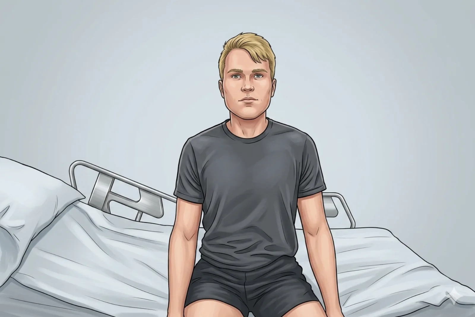
- Patient seated; arm relaxed at side
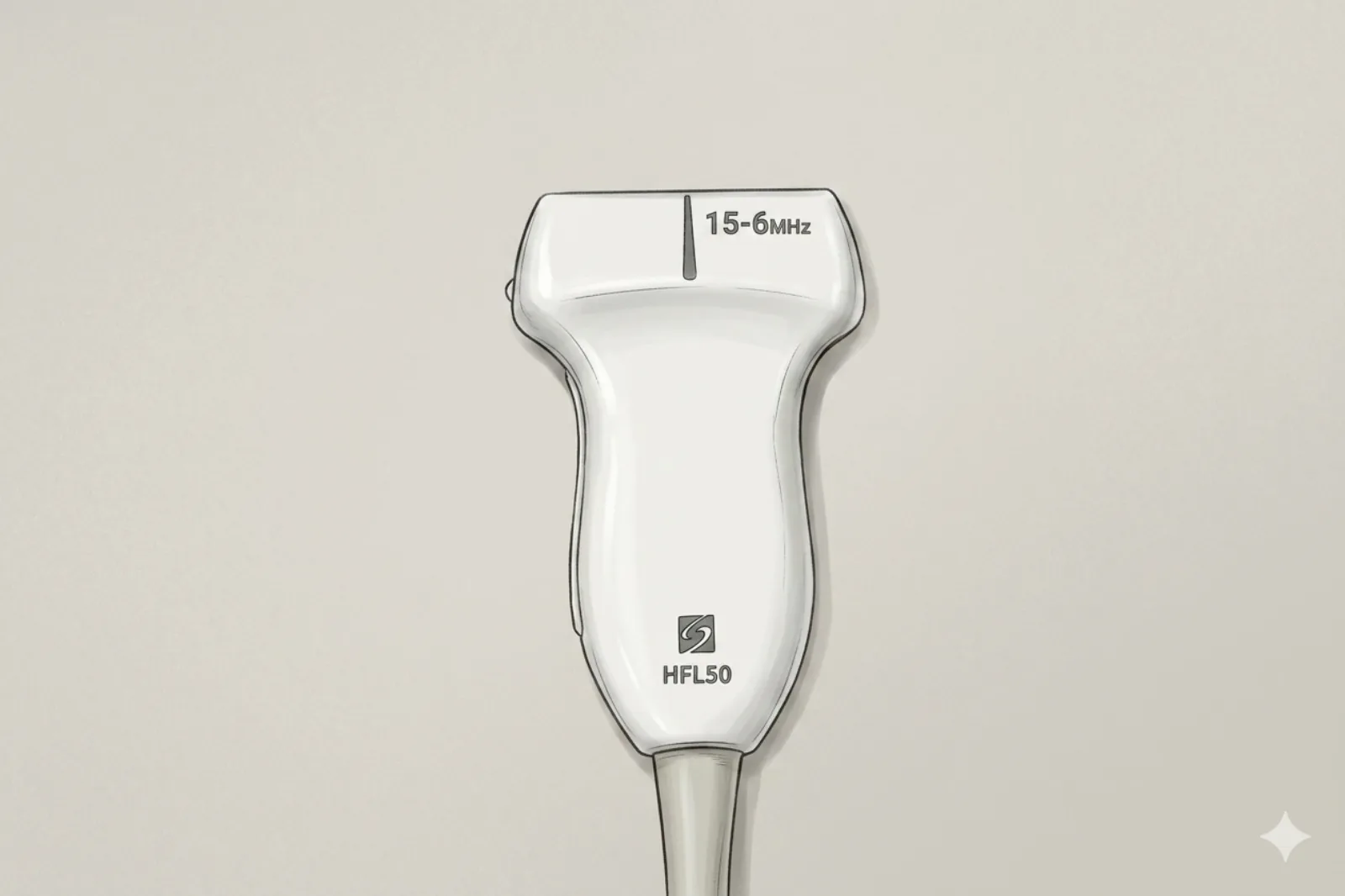
Probe
- High-frequency linear probe (10-15 MHz)
Landmarking — find the view
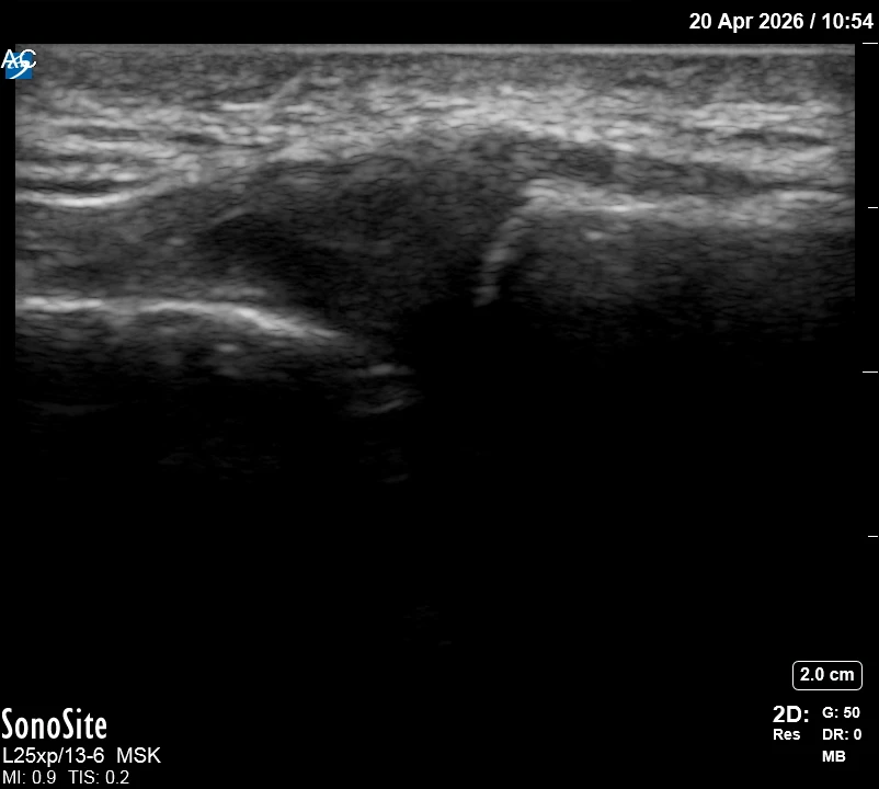
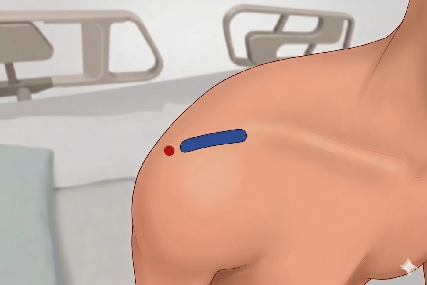
- Start mid-clavicle; slide laterally to the gap; continue to acromion; translate to center the joint gap
The correct ultrasound view
- Two flat bones (clavicle/acromion) with thin dark joint line centered
Needle corridor
- Superior approach: between superior capsule (below) and subcutaneous tissue (above) → tiny joint space, angled to match the oblique joint plane
Avoid
- Acromial branch of thoracoacromial artery
- Overfilling (very small capacity)
Steps
- 1In-plane SUPERIOR approach along the oblique joint plane; angle the needle to match the joint line rather than the skin surface.
- 2Confirm trajectory by tilting probe to reveal the joint cleft orientation before entry.
- 3Inject 0.5-1 mL total; stop if resistance or pain rises.
- 4STOP IMMEDIATELY if the patient reports electric, shooting, or paresthetic sensations.
Tips
- Use minimal local anesthetic to save space for steroid.
- Limit corticosteroid to ≤ 3 per joint per year.
Resident pearls
- Don't 'fill up' the joint with local if you plan to use steroid; the capacity is often < 1 mL.
- Total injectate volume ≤ 1 mL — pre-mix steroid and small lidocaine aliquot in the same syringe before approaching the joint.
- If pressure is low, the capsule may be incompetent (post-trauma/surgery), leaking into SA space.
- Look for the Geyser sign: fluid tracking from GH joint up through the AC joint.
Supplies
- 3 mL Syringe
- 25G 1.5" Needle (default); 27G 1" only for lean patients — 27G deflects before reaching the joint in larger patients
- 18G Drawing Needle
- 1% Lidocaine (0.5 mL)
- Triamcinolone 20-40 mg OR Dexamethasone 4 mg (0.25-0.5 mL)
- Alcohol Swabs (vial tops)
- Betadine/Chloraprep
- Sterile Gloves
- Sterile 4x4 Gauze Pads
- Sterile Towels & Drapes
- Adhesive Bandage (Band-Aid)
- Washcloth/Towel (gel cleanup)
- Needle Disposal Safety Cap
- Sterile probe cover & gel
Post-procedure
- Apply a clean dressing; remove after 24 hours.
- Local anesthetic relief for 1-4 hours; steroid begins 24-72 hours.
- Steroid 'flare' in 2–10% — manage with ice and acetaminophen.
- Rest 24-48 hours.
- Return immediately for: spreading redness, swelling, fever, or disproportionate pain.
- Diabetics: expect 50-100 mg/dL glucose rise for 3-5 days.
- Maximum 3 injections per joint per year.