Ultrasound · Shoulder
Long Head of the Biceps Tendon Sheath Injection
shoulderbicepssheathtenosynovitis
Treats
- Biceps tendon sheath inflammation
- Diagnostic sheath injection
Contraindications
- Active local skin infection at entry site (absolute)
- Suspected septic tenosynovitis (absolute)
- Known allergy to local anesthetic, steroid, or antiseptic prep
- Therapeutic anticoagulation — review case-by-case
- Uncontrolled diabetes (relative)
- Recent biceps tendon rupture or high-grade tear (relative — particulate steroid increases re-rupture risk)
Pre-procedure checklist
- Confirm patient identity (two identifiers)
- Confirm procedure, side, and site
- Review allergies and anticoagulation
- Confirm informed consent
- Pre-scan with Doppler to identify the AHC artery position in the groove
- Time-out immediately before needle entry
Positioning
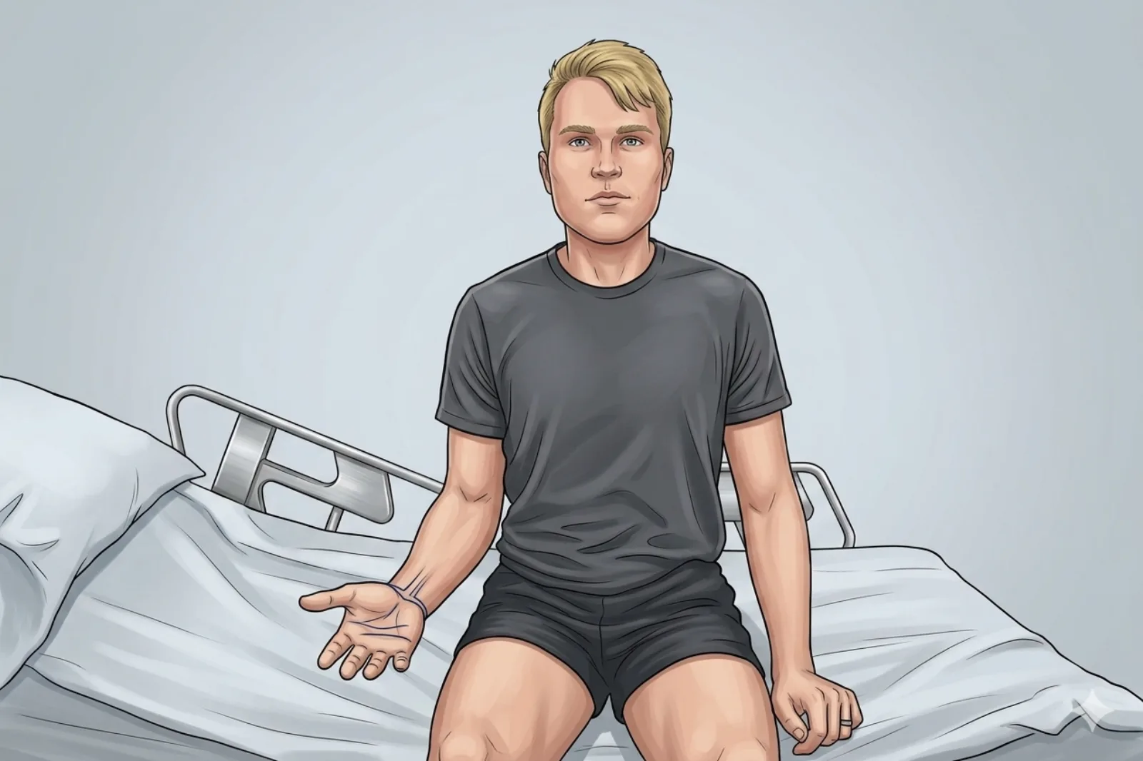
- Patient seated; forearm supinated and resting on the lap or a table (palm up)
- Slight external rotation of the shoulder brings the bicipital groove anterior and accessible
Probe
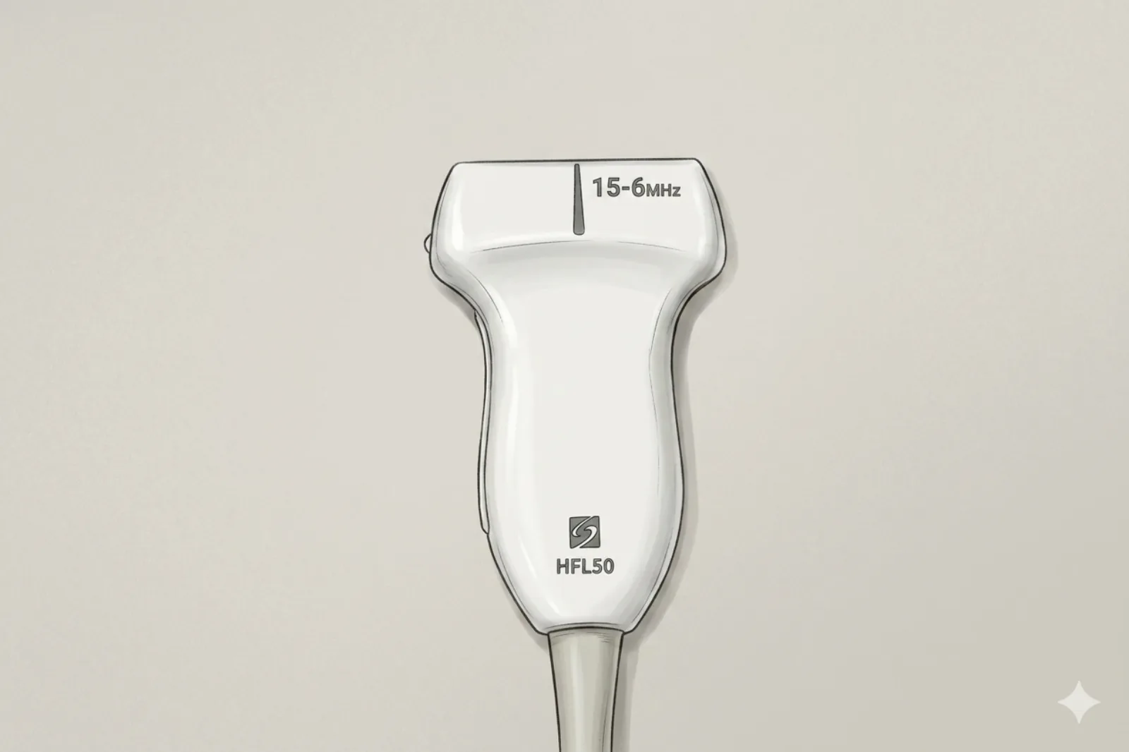
- High-frequency linear probe (6-13 MHz) (short footprint helpful)
Landmarking — find the view
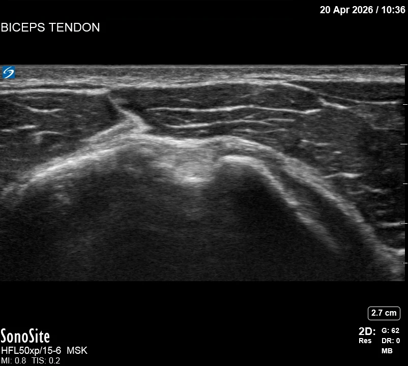
- Visual Cue: The "Bicipital Valley" — identify the deep bony groove between the greater and lesser tuberosities.
- Center the round, hyperechoic biceps tendon within the groove.
- Rotate 90° into long-axis to confirm the tendon is intact and not subluxed.
- Doppler Survey: Identify the ascending branch of the anterior humeral circumflex artery (typically transverse across the groove, often visualized medial in the groove near the lesser tuberosity).
The correct ultrasound view
- Round/oval tendon centered in groove; thin dark rim around it = sheath
Needle corridor
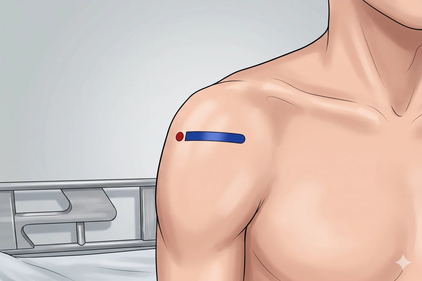
- Lateral → medial path (from greater tuberosity side) into the thin dark rim around tendon — stop the tip in the sheath BEFORE reaching the medial groove wall where the AHC artery typically lies. Not intratendinous.
Avoid
- Ascending branch of the anterior humeral circumflex artery (Doppler before every entry — if it sits in the planned path, re-plan)
- Intratendinous steroid
Steps
- 1Advance in-plane lateral-to-medial; inject a tiny test amount for circumferential 'target sign'; inject the planned volume once confirmed.
- 2STOP IMMEDIATELY if the patient reports electric, shooting, or paresthetic sensations.
Tips
- If fluid collects above tendon or into SA space, you are in the wrong layer — readjust before steroid.
- The biceps sheath communicates with the GH joint — anticipate some intra-articular steroid spread. Lower doses reduce systemic load and rupture risk.
- Limit to ≤ 3 injections per year per tendon.
Resident pearls
- Create a "test effusion" with local to confirm circumferential spread around the tendon.
- Always Doppler before entry — the AHC artery position varies; if it sits in the planned path, re-plan.
- Total injectate (anesthetic + steroid) ≤ 3 mL to avoid overfilling the sheath and retrograde GH joint tracking.
Supplies
- 5 mL Syringe
- 25G 1.5" Needle
- 18G Drawing Needle
- 1% Lidocaine (1-2 mL)
- Triamcinolone 20-40 mg OR Dexamethasone 4-8 mg
- Alcohol Swabs (vial tops)
- Betadine/Chloraprep
- Sterile Gloves
- Sterile 4x4 Gauze Pads
- Sterile Towels & Drapes
- Adhesive Bandage (Band-Aid)
- Washcloth/Towel (gel cleanup)
- Needle Disposal Safety Cap
- Sterile probe cover & gel
Post-procedure
- Apply a clean dressing; remove after 24 hours.
- Local anesthetic relief for 1-4 hours; steroid begins 24-72 hours.
- Steroid 'flare' in 2–10% — manage with ice and acetaminophen.
- Rest the shoulder 24-48 hours; avoid heavy lifting for 1 week.
- Return immediately for: spreading redness, swelling, fever, or disproportionate pain.
- Diabetics: expect 50-100 mg/dL glucose rise for 3-5 days.
- Maximum 3 injections per tendon per year.