Ultrasound · Hand
Carpal Tunnel Injection
wristcarpal tunnelmedian nerve
Treats
- Median nerve compression (carpal tunnel syndrome)
Contraindications
- Active local skin infection at entry site (absolute)
- Suspected septic compartment (absolute)
- Known allergy to local anesthetic, steroid, or antiseptic prep
- Therapeutic anticoagulation — review case-by-case (closed-compartment hematoma risk)
- Persistent median artery in the planned corridor (relative — re-plan or defer)
- Severe denervation atrophy with thenar wasting (relative — refer for surgical release)
- Uncontrolled diabetes (relative)
Pre-procedure checklist
- Confirm patient identity (two identifiers)
- Confirm procedure, side, and site — mark with patient awake
- Review allergies and anticoagulation
- Confirm informed consent
- Pre-scan: identify median nerve, ulnar bundle, and Doppler-screen for persistent median artery and bifid nerve variant
- Time-out immediately before needle entry
Positioning
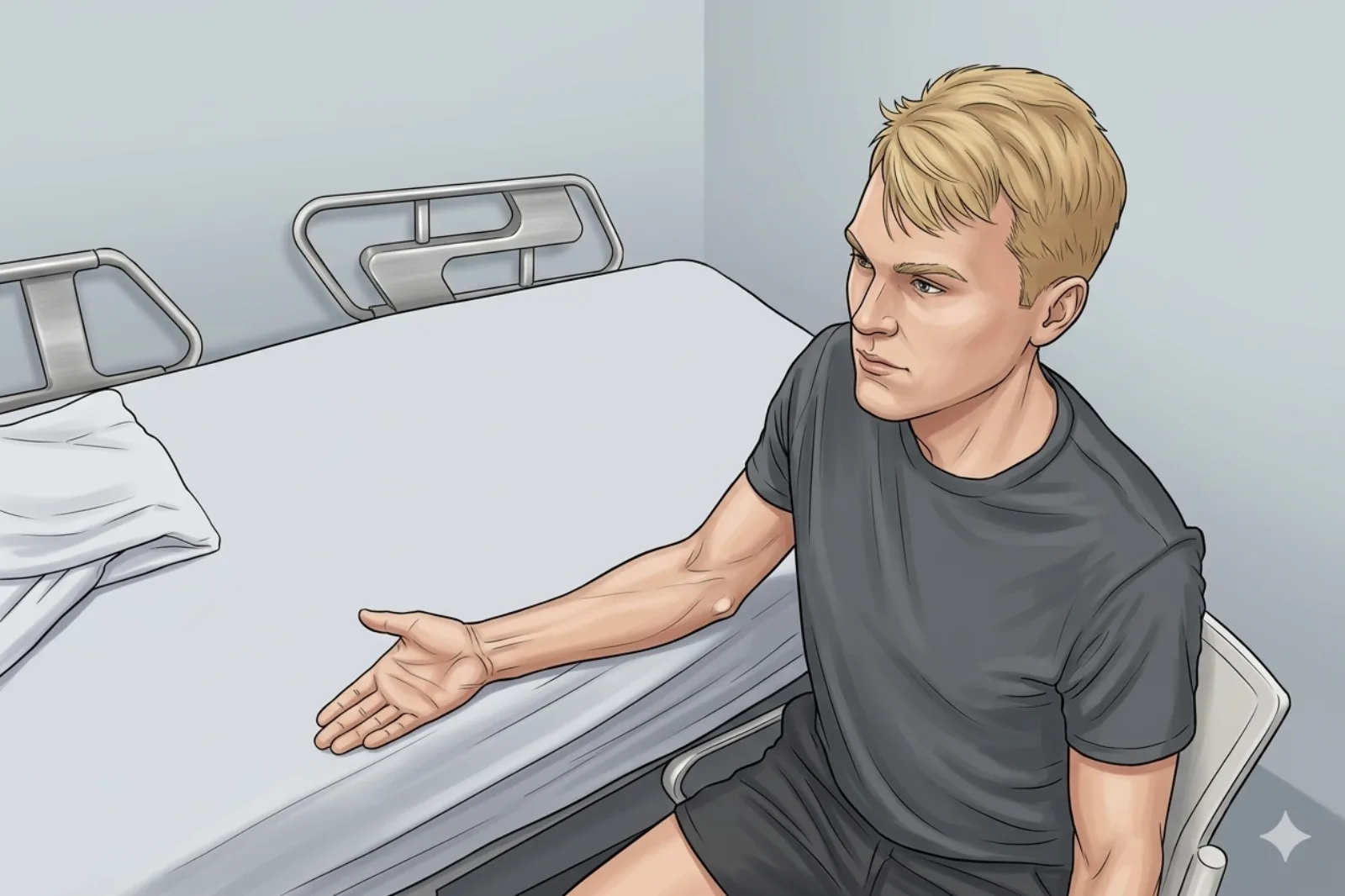
- Seated preferred (or supine if vasovagal); forearm supinated; wrist slightly extended
Probe
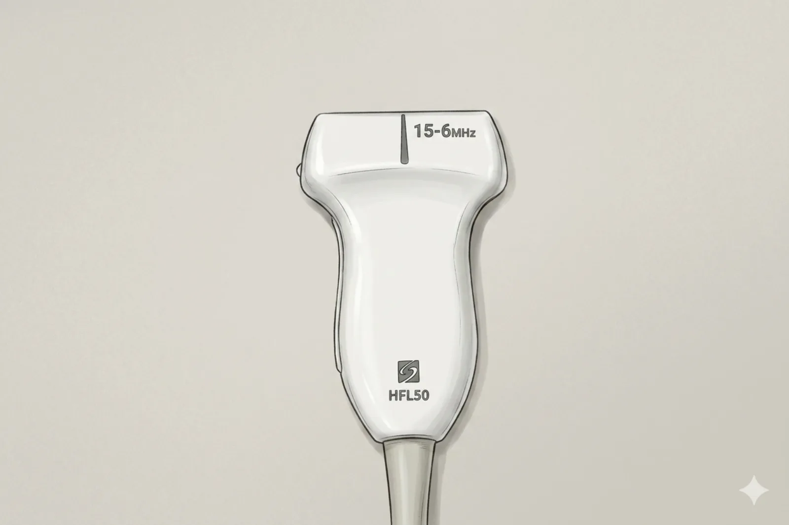
- High-frequency linear probe (15–18 MHz); full footprint allows visualization of the entire carpal tunnel in a single view
Landmarking — find the view
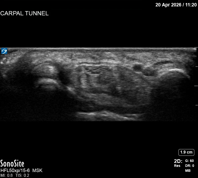
- Probe transversely at proximal wrist crease → identify median nerve under retinaculum → identify flexor tendons → map ulnar bundle → check for bifid nerve/persistent median artery
The correct ultrasound view
- Median nerve centered; retinaculum as bright roof; adequate perineural space
Needle corridor
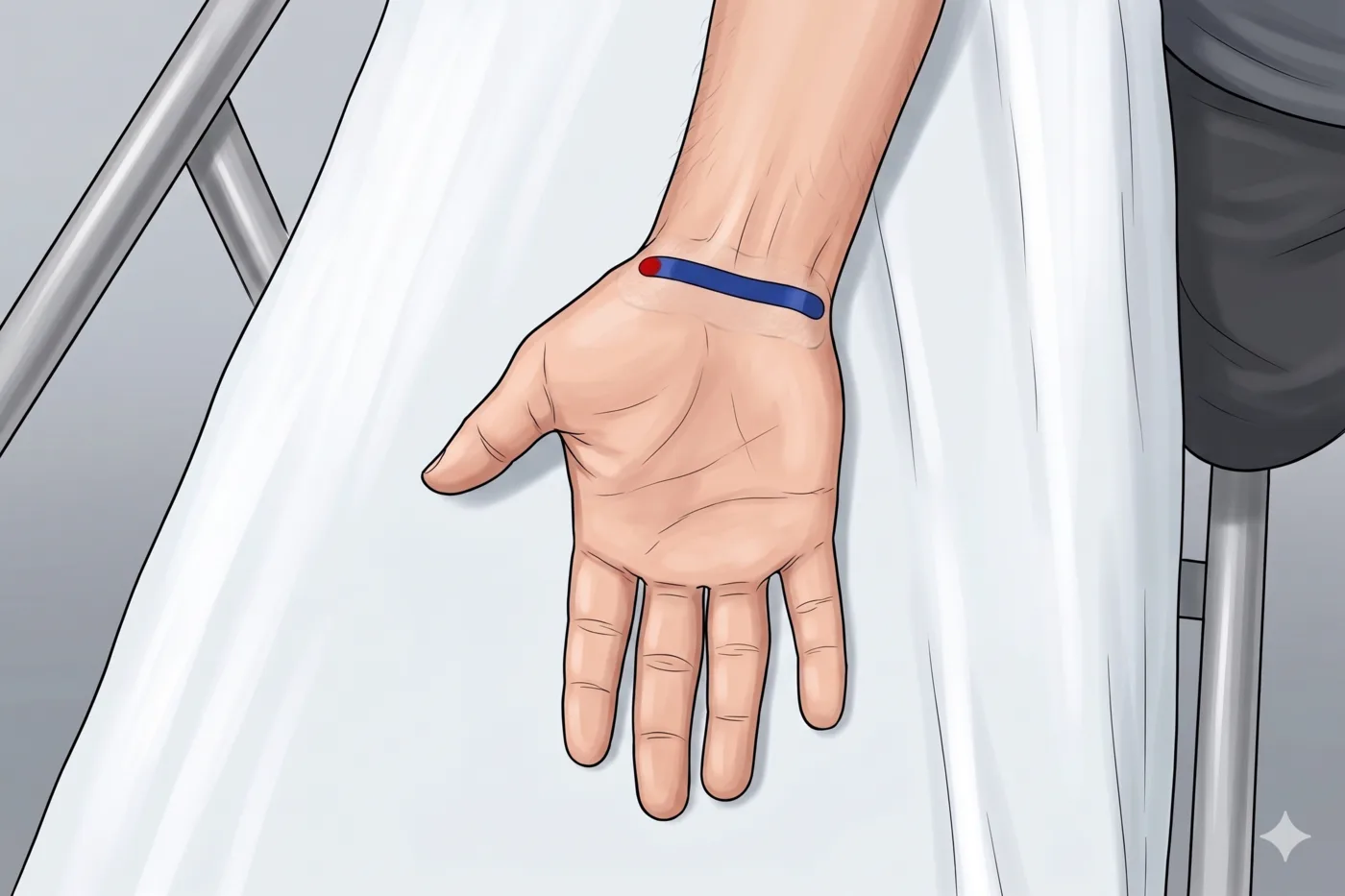
- In-plane ulnar entry (ulnar to the ulnar artery and nerve — map both first). Advance the needle tip to lie immediately ulnar to the median nerve, deep to the flexor retinaculum. DO NOT cross the tip past the nerve to the radial side.
Avoid
- Median nerve — identify and keep the needle tip ulnar to the nerve at all times; never inject into the nerve substance
- Ulnar nerve/artery
- Persistent median artery (if in path: ABANDON, re-plan, or defer)
- Bifid median nerve variant — injecting between halves risks arterial laceration
- Palmar cutaneous branch of the median nerve — arises proximal to the tunnel and runs superficially toward the thenar skin; map before entry
- Superficial palmar arch — lies distal to the tunnel; keep the target proximal to the arch
Steps
- 1In-plane ulnar entry; position the tip immediately ulnar to the median nerve, deep to retinaculum.
- 2Inject 1-2 mL slowly, hydrodissecting the ulnar margin of the nerve — watch for circumferential 'target sign' spread.
- 3STOP IMMEDIATELY if the patient reports electric, shooting, or paresthetic sensations into the hand or fingers — withdraw 1-2 mm and reposition before any further injection.
Tips
- Use low volume — total injectate ≤ 3 mL to avoid further median nerve compression in this tight compartment.
- Confirm small fluid rings around nerve — do not advance the needle tip past the nerve to the radial side.
- If color Doppler shows a persistent median artery in the planned path, ABANDON the ulnar approach and either use a different angle or defer the procedure.
Resident pearls
- The “Oven Mitt” Rule: The flexor retinaculum is a tight mitt. Slide the needle UNDER the mitt, not into the hand (nerve).
- Target Sign: Create a circumferential halo by hydrodissecting the ulnar margin of the nerve.
- Persistent median artery + bifid median nerve occurs in 5-10%. Injection between the two halves can lacerate the artery and cause hematoma in a closed compartment — re-plan or defer.
- The ulnar approach (medial-to-lateral) is safest to avoid the radial neurovascular bundle — but stop the tip ulnar to the median nerve.
Supplies
- 3 mL Syringe
- 25G or 27G 1" Needle (1.5" is unnecessarily long for this superficial target)
- 18G Drawing Needle
- 1% Lidocaine (1-2 mL)
- Dexamethasone 4-10 mg (PREFERRED — non-particulate, safer near nerve) OR methylprednisolone 40 mg
- Alcohol Swabs (vial tops)
- Betadine/Chloraprep
- Sterile Gloves
- Sterile 4x4 Gauze Pads
- Sterile Towels & Drapes
- Adhesive Bandage (Band-Aid)
- Washcloth/Towel (gel cleanup)
- Needle Disposal Safety Cap
- Sterile probe cover & gel
Post-procedure
- Apply a clean dressing; remove after 24 hours.
- Local anesthetic relief for 1-4 hours; steroid begins 24-72 hours.
- Steroid 'flare' in 2–10% — manage with ice and acetaminophen.
- Rest the wrist 24-48 hours; resume normal activity as tolerated thereafter.
- Return immediately for: spreading redness, swelling, fever, disproportionate pain, OR new/worsening numbness or weakness.
- Diabetics: expect 50-100 mg/dL glucose rise for 3-5 days.
- Maximum 3 injections per year. If symptoms recur quickly, discuss surgical release.