Ultrasound · Hand
First Dorsal Compartment Injection (De Quervain's)
wristde quervainaplepb
Treats
- De Quervain's tenosynovitis (APL/EPB)
Contraindications
- Active local skin infection at entry site (absolute)
- Suspected septic tenosynovitis (absolute)
- Known allergy to local anesthetic, steroid, or antiseptic prep
- Therapeutic anticoagulation — review case-by-case
- Uncontrolled diabetes (relative)
- Prior steroid injection at this site within 3 months (relative — fat atrophy/depigmentation cumulative risk)
Pre-procedure checklist
- Confirm patient identity (two identifiers)
- Confirm procedure, side, and site
- Review allergies and anticoagulation
- Confirm informed consent — DISCUSS subcutaneous fat atrophy and skin depigmentation risk at the radial styloid
- Pre-scan: identify APL/EPB tendons, septum (if present), superficial radial nerve branches, radial artery
- Time-out immediately before needle entry
Positioning
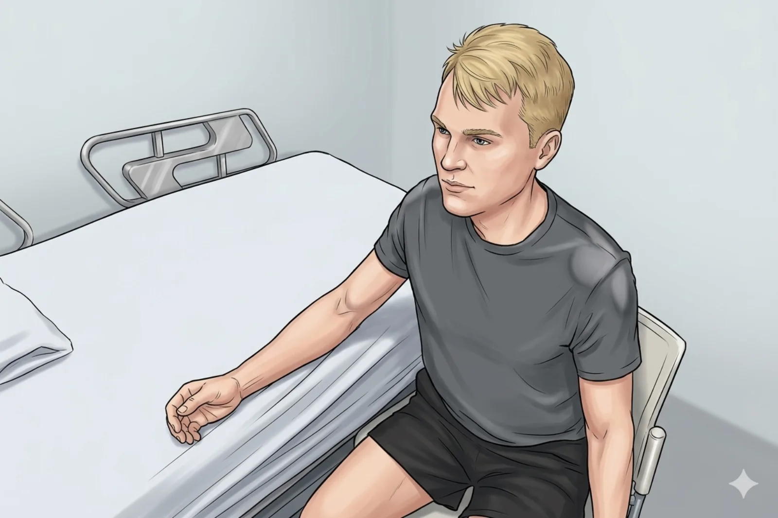
- Seated or supine; forearm neutral; thumb up
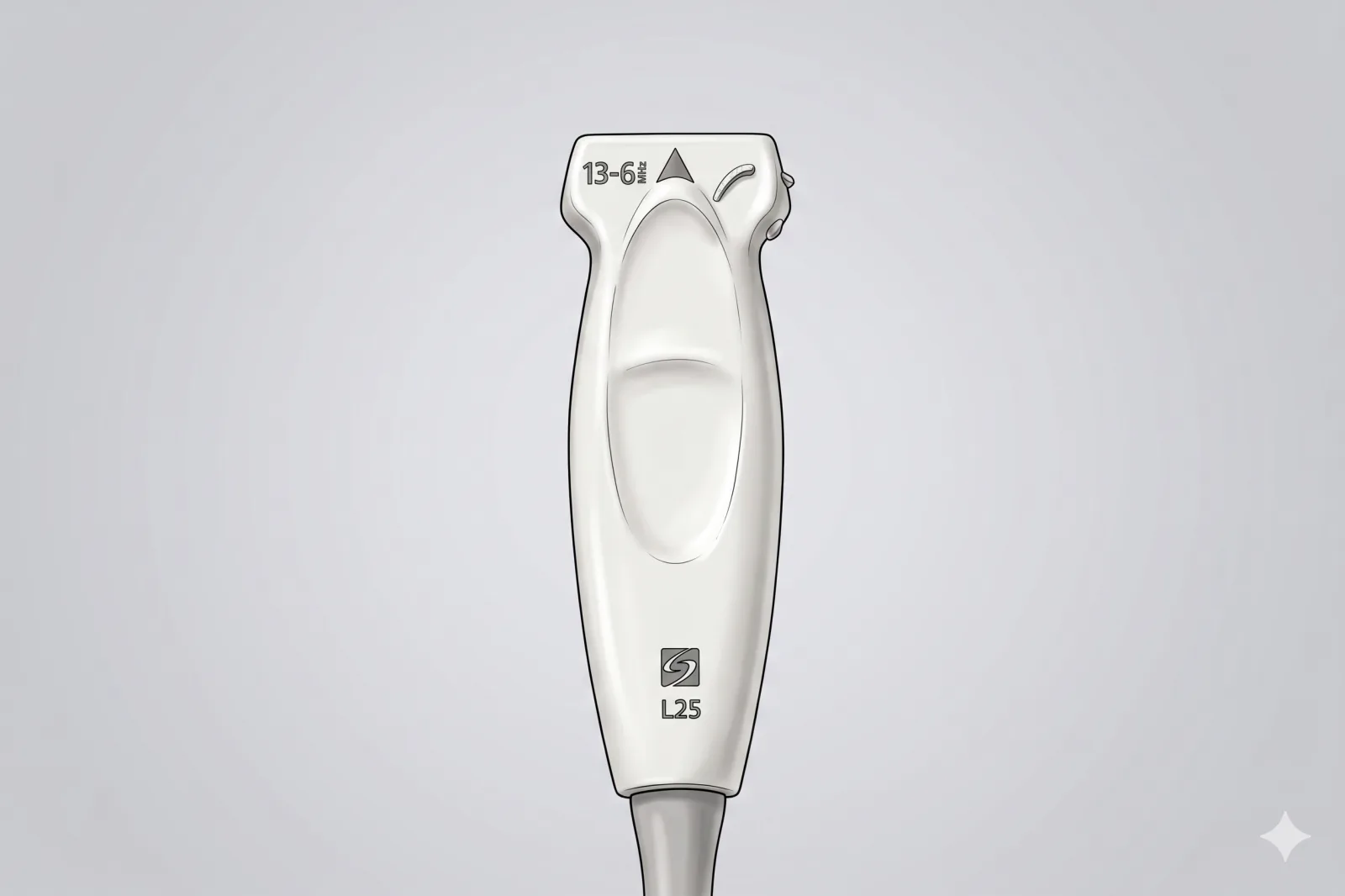
Probe
- High-frequency linear probe (6-13 MHz)
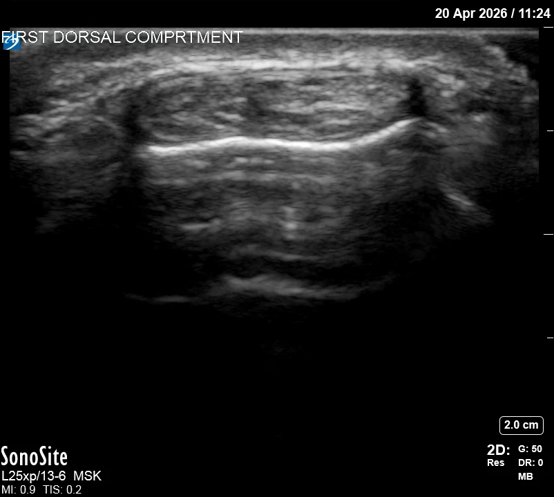
Landmarking — find the view
- Visual Cue: 'Two eyes in one socket' — identify the APL and EPB tendons together in short-axis over the radial styloid.
- Slide the probe slightly proximal and distal to identify any vertical septum (a wall) between the tendons.
- Check for any small fluid collections or 'halo' around the pair.
- Map the superficial radial nerve branches — they are frequently very close to the entry site.
- Identify the radial artery on the volar side — DO NOT use a volar trajectory.
The correct ultrasound view
- Two tendons within sheath; septum visible if present
Needle corridor
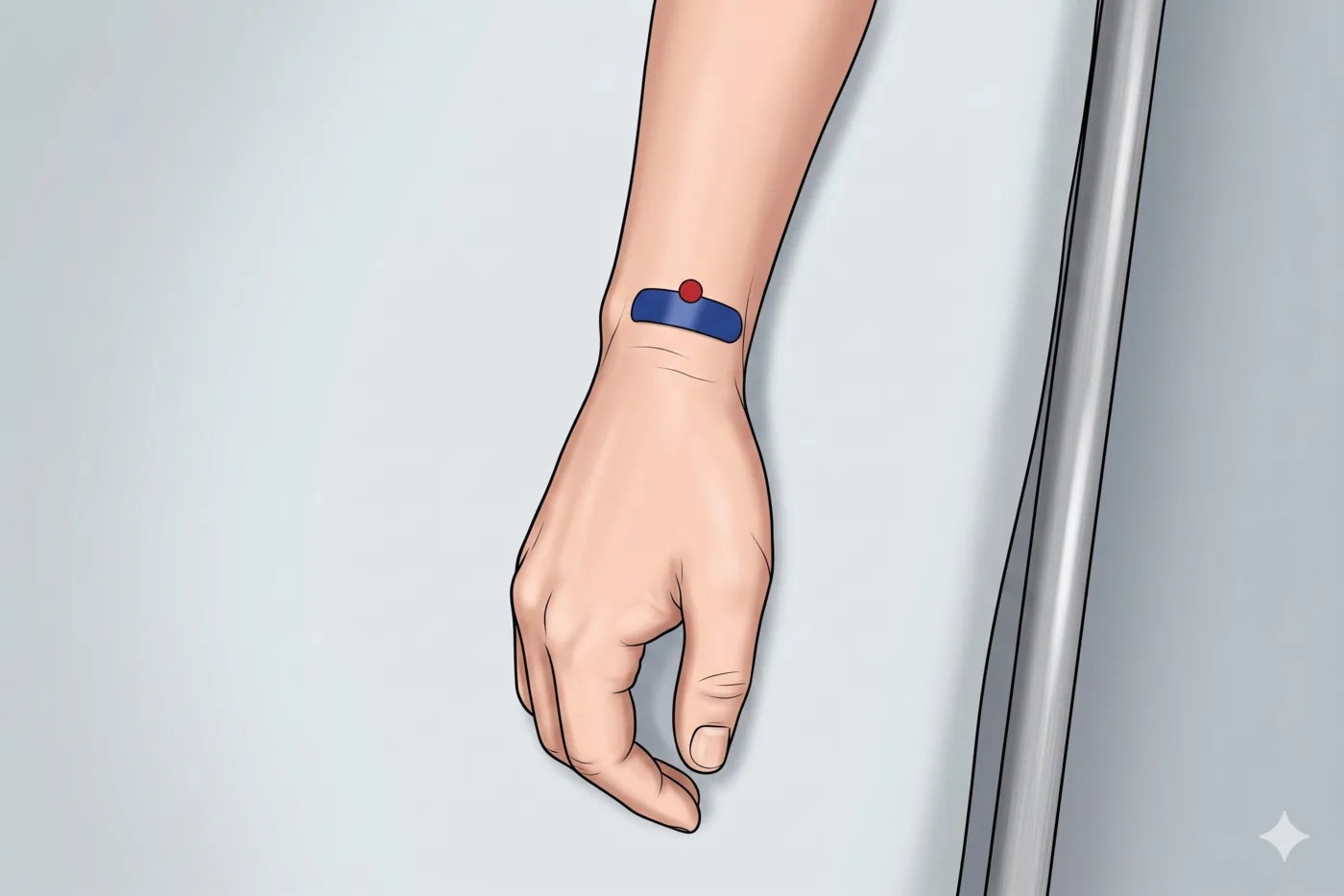
- Dorsal in-plane approach (after rotating the probe into long-axis along the tendons): enter from the dorsal/dorsoradial aspect distal-to-proximal, advancing into the sheath superficial to the APL/EPB tendons. AVOID any volar trajectory — the radial artery lies on the volar side of the radial styloid.
Avoid
- Superficial radial nerve branches
- Radial artery (volar — never use a volar approach)
- Intratendinous injection
Steps
- 1Dorsal in-plane entry into the sheath; inject ≤ 1.5 mL total; ensure spread around BOTH tendons.
- 2If a septum is present, perform TWO separate injections — one in the APL sub-sheath and one in the EPB sub-sheath. A single injection into the APL alone is the most common cause of treatment failure.
- 3STOP IMMEDIATELY if the patient reports electric, shooting, or paresthetic sensations along the dorsoradial hand or thumb.
Tips
- Always identify the superficial radial nerve first and adjust approach.
- Use a short 27G needle for better control in this superficial space — but only with non-particulate steroid (27G can clog with triamcinolone suspension).
- Counsel patient about subcutaneous fat atrophy and depigmentation risk at the radial styloid (well-documented particulate steroid complication).
- Limit to 1-2 steroid injections per year at this site.
Resident pearls
- EPB sub-compartmentalization (vertical septum) is present in 20-60% of symptomatic cases and is THE single most common cause of treatment failure — actively seek it and inject both sub-sheaths.
- Wrist in neutral to slight ulnar deviation avoids anisotropy artifacts.
Supplies
- 3 mL Syringe
- 27G 0.5" or 1" Needle (use ≥ 25G if injecting triamcinolone — particulate can clog 27G)
- 18G Drawing Needle
- 1% Lidocaine (0.5-1 mL)
- Dexamethasone 4 mg (PREFERRED — non-particulate, no fat atrophy risk near SRN) OR triamcinolone 10-20 mg
- Alcohol Swabs (vial tops)
- Betadine/Chloraprep
- Sterile Gloves
- Sterile 4x4 Gauze Pads
- Sterile Towels & Drapes
- Adhesive Bandage (Band-Aid)
- Washcloth/Towel (gel cleanup)
- Needle Disposal Safety Cap
- Sterile probe cover & gel
Post-procedure
- Apply a clean dressing; remove after 24 hours.
- Local anesthetic relief for 1-4 hours; steroid begins 24-72 hours.
- Steroid 'flare' in 2–10% — manage with ice and acetaminophen.
- Rest the thumb 24-48 hours; brief thumb spica splint optional for 1 week.
- Return for: spreading redness, swelling, fever, disproportionate pain, OR new skin depigmentation/dimpling at the injection site.
- Diabetics: expect 50-100 mg/dL glucose rise for 3-5 days.
- Maximum 1-2 injections per year at this site.