Before You Listen
- Prerequisites: CVA-01 vascular anatomy and stroke syndromes (Circle of Willis, MCA face-and-arm pattern, lacunar localization); the basic difference between ischemic and hemorrhagic stroke; familiarity with the Glasgow Coma Scale (GCS) and basic ICU pharmacology of intravenous (IV) antihypertensives (labetalol, nicardipine).
- Runtime: 49 minutes.
- Topic in one line: the five-subtype Trial of Org 10172 in Acute Stroke Treatment (TOAST) classification and the secondary prevention it drives, the National Institutes of Health Stroke Scale (NIHSS) zero-to-42 severity tool with its 17%-per-point outcome gradient and posterior-circulation blind spot, the modified Rankin Scale (mRS) with functional independence defined as 0-2, the alteplase 0.9 mg/kg / 90 mg cap / 10% bolus / 60-minute infusion protocol with the NINDS 0-3 hour and ECASS III 3-4.5 hour windows and pooled numbers needed to treat (NNT) of about 10 and 20, the NINDS 6.4% symptomatic intracerebral hemorrhage (sICH) rate, the 185/110 pre-treatment and 180/105 post-treatment blood-pressure thresholds, glucose as the lab that must result and the ≤45-minute door-to-needle goal, the original ECASS III combined diabetes-plus-prior-stroke exclusion, tenecteplase (TNK) 0.25 mg/kg / 25 mg cap as a single bolus, the WAKE-UP DWI-FLAIR mismatch and EXTEND perfusion mismatch trials, and the Class of 2015 thrombectomy trials with DAWN clinical-core mismatch out to 24 hours and DEFUSE 3 perfusion mismatch out to 16 hours, plus the conventional ASPECTS ≥6 cutpoint. Memorize them cold, and keep trial enrollment rules distinct from current clinical eligibility.
Vignette. A 74-year-old right-handed woman with atrial fibrillation (not on anticoagulation), diabetes, and well-controlled hypertension is brought to a primary stroke center 95 minutes after the sudden onset of right facial droop, dense right-arm flaccidity, and global aphasia witnessed by her husband. Her blood pressure on arrival is 192/108. Initial fingerstick glucose is 118 mg/dL. Non-contrast head computed tomography (CT) is unremarkable for hemorrhage with an Alberta Stroke Program Early CT Score (ASPECTS) of 9; CT angiography (CTA) shows occlusion of the proximal left middle cerebral artery (MCA) at the M1 segment. National Institutes of Health Stroke Scale (NIHSS) is 18. The comprehensive stroke center is 35 minutes away by helicopter.
What is the next pharmacologic step and the exact dose, what blood-pressure target must be achieved before that drug is given and maintained after, what is the only mandatory pre-treatment laboratory test, and what is the rationale for proceeding to mechanical thrombectomy in parallel rather than waiting to see if the drug works?
(Answer at the end of this chapter)
Section 1: TOAST Classification, NIHSS, and the Modified Rankin Scale
Bottom line: the TOAST classification sorts ischemic stroke into five etiologic subtypes that drive secondary prevention, and the workup never delays reperfusion; the National Institutes of Health Stroke Scale (NIHSS) is a 0-42 acute-severity tool that under-weights posterior strokes, and each point carried 17% lower odds of an excellent outcome in the TOAST cohort; the modified Rankin Scale (mRS) is the global disability scale, with functional independence = mRS 0-2.
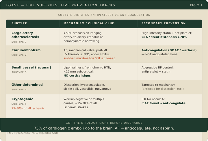
Every acute ischemic stroke is sorted by mechanism because the secondary prevention regimen depends on it. Do that workup alongside acute care, never in front of it: mechanism decides prevention, not whether to reperfuse. The Trial of Org 10172 in Acute Stroke Treatment (TOAST) classification organizes ischemic strokes into five etiologic subtypes. Large artery atherosclerosis captures strokes from atheromatous plaques in the internal carotid artery, vertebral artery, or major intracranial vessels. The plaque either causes hemodynamic narrowing or seeds an artery-to-artery embolus; vascular imaging typically reveals greater than 50% stenosis. Secondary prevention combines a high-intensity statin, antiplatelet therapy, and vascular-risk control. Carotid endarterectomy or stenting is reserved for selected symptomatic extracranial carotid disease, conventionally severe stenosis of at least 70%, after assessment of surgical candidacy; a stenotic artery alone does not book the operating room, and intracranial stenting is not first-line. Cardioembolism is the second subtype; the heart harbors the clot and the brain catches the embolus. The most common cardiac source is atrial fibrillation, with mechanical valves, recent myocardial infarction with left ventricular thrombus, dilated cardiomyopathy, endocarditis, and patent foramen ovale rounding out the list. The clinical clue is a sudden maximal deficit at onset (no stuttering progression). Atrial fibrillation-associated stroke is prevented with anticoagulation, not antiplatelet therapy alone, unless anticoagulation is contraindicated. The rest of the cardioembolic list is managed by source: endocarditis, a mechanical valve and a patent foramen ovale each carry their own regimen, and embolic stroke of undetermined source does not get empiric anticoagulation. AHA/ASA secondary-prevention summary.
Small vessel occlusion (lacunar stroke) is the third subtype, covered fully in CVA-01: lipohyalinosis of small penetrators from chronic hypertension produces a less than 15 mm subcortical infarct in the basal ganglia, thalamus, internal capsule, or pons, with no cortical signs; secondary prevention is antiplatelet therapy, a statin and vascular-risk control, with blood pressure the dominant target. Stroke of other determined etiology is the wildcard subtype: arterial dissection, hypercoagulable states, sickle cell disease, vasculitis, moyamoya. Stroke of undetermined etiology (cryptogenic) applies when the workup fails to identify a definitive cause, when multiple causes are found simultaneously, or when the workup is incomplete, and accounts for approximately 25-30% of all ischemic strokes. Prolonged cardiac monitoring with implantable loop recorders has revealed occult atrial fibrillation in a substantial proportion of cryptogenic patients, shifting many into the cardioembolic category and from antiplatelet therapy to anticoagulation.
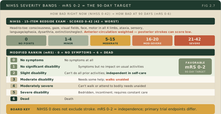
The National Institutes of Health Stroke Scale (NIHSS) is the standardized 15-item acute-severity tool, scored 0-42. Items cover level of consciousness, orientation questions, response to commands, horizontal eye movements, visual fields, facial symmetry, motor function in each limb, limb coordination, sensation, language (naming, reading, describing), dysarthria, and extinction/inattention. Severity cutpoints: 1-4 minor, 5-15 moderate, 16-20 moderate-to-severe, 21-42 severe. A score above 20 is the conventional clue to a large vessel occlusion (LVO), and vascular imaging is what confirms one; a low score does not exclude an LVO. A score above 25 is associated with death or major disability, and it neither fixes an individual patient’s outcome nor by itself prohibits treatment. In the 1,281-patient TOAST cohort, each additional NIHSS point carried approximately 17% lower odds of an excellent GOS/Barthel outcome at three months. Every point matters, and that figure is a cohort association rather than a per-patient forecast. Adams et al., 1999.
The most critical board-tested limitation of the NIHSS is that it under-represents posterior circulation strokes. The scale is heavily weighted toward anterior-circulation deficits (language, neglect, gaze, facial strength, limb motor function are all prominent in MCA strokes). A patient with a devastating basilar occlusion causing vertigo, severe dysphagia, bilateral unsteadiness, and cranial nerve deficits can score deceptively low because those posterior-fossa deficits are poorly captured. The NIHSS also does not test memory, executive function, emotional state, or swallowing. A patient with a thalamic stroke causing severe amnesia may score 0 despite a devastating cognitive deficit; a patient with a lateral medullary stroke and severe dysphagia at high risk for aspiration may score 3 or 4. The NIHSS measures acute severity, not total functional burden.
The modified Rankin Scale (mRS) is the primary outcome measure used in stroke clinical trials. It rates global disability on a 7-point scale: 0 = no symptoms; 1 = no significant disability (some symptoms but all usual activities preserved); 2 = slight disability (cannot do all previous activities but independent in self-care); 3 = moderate disability (requires some help, can walk without assistance); 4 = moderately severe (cannot walk and cannot attend to bodily needs without assistance); 5 = severe (bedridden, incontinent, constant nursing care); 6 = death. Functional independence = mRS 0, 1, or 2 at 90 days. When a board question references functional independence in the context of a stroke trial, it means a 90-day mRS of 2 or below. That definition is not the same as every trial’s primary endpoint: ECASS III used mRS 0-1, NINDS used a global four-scale outcome, MR CLEAN, SWIFT PRIME, REVASCAT and DEFUSE 3 analyzed the ordinal mRS distribution, and EXTEND-IA used early reperfusion and day-3 neurologic improvement as coprimary outcomes.
High Yield — TOAST, NIHSS, and mRS
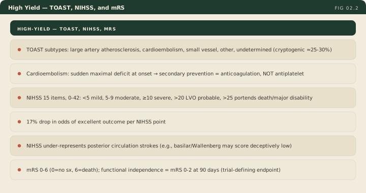
- TOAST subtypes: large artery atherosclerosis, cardioembolism, small vessel (lacunar), other determined etiology, undetermined (cryptogenic ≈ 25-30%).
- Cardioembolism clue: sudden maximal deficit at onset; AF-associated stroke is prevented with anticoagulation (NOT antiplatelet alone). Other cardiac sources are managed source by source.
- NIHSS = 15 items, 0-42; 1-4 minor, 5-15 moderate, 16-20 moderate-to-severe, 21-42 severe; >20 is the conventional LVO clue (imaging confirms it), >25 is associated with death/major disability.
- 17% lower odds of an excellent outcome per NIHSS point (TOAST cohort, GOS/Barthel at three months).
- NIHSS under-represents posterior circulation strokes: a basilar/Wallenberg patient may score deceptively low.
- mRS 0-6: 0 = no symptoms; 6 = death.
- Functional independence = mRS 0-2 at 90 days. Primary endpoints differ by trial: ECASS III used mRS 0-1, and several EVT trials used the ordinal mRS shift.
- Cryptogenic stroke: implantable loop recorder reveals occult AF in a substantial fraction; reclassification shifts secondary prevention to anticoagulation.
Clinical Pearl — 75% of cardiogenic emboli go to the brain
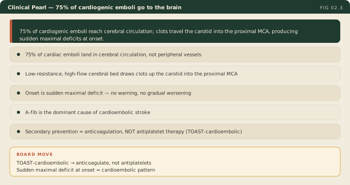
Roughly 75% of clots launched from the heart end up in the cerebral circulation rather than in peripheral vessels. The cerebral arterial bed is a low-resistance high-flow target, and emboli that travel up the carotid take the path of least resistance into the proximal MCA. That tropism is why the heart is the first organ you interrogate after a sudden maximal cortical deficit. It is an anatomic association, not an argument that atrial fibrillation is common, and it does not license one prevention regimen for the whole cardioembolic category: AF-associated stroke gets anticoagulation, and the other sources get source-specific therapy.
The thalamus is a critical relay center. A stroke there can cause devastating severe amnesia. The patient cannot form a single new memory.
— CVA-02 podcast, ~12:46
So a patient with a small vessel occlusion in the internal capsule might have dense hemiplegia, but they won’t have aphasia. They won’t have visual neglect.
— CVA-02 podcast, ~6:23