Before You Listen
- Prerequisites: vascular anatomy of the anterior cerebral artery (ACA), middle cerebral artery (MCA) and posterior cerebral artery (PCA) territories from CVA-01; the watershed (border-zone) concept from CVA-01; basic disorders-of-consciousness terminology; the median-nerve somatosensory evoked potential (SSEP) waveform from electrodiagnostics.
- Runtime: approximately 54 minutes 50 seconds including the opening promo.
- Topic in one line: the global brain-oxygenation event. Definitions of anoxia, hypoxia and ischemia and the global-versus-focal contrast with stroke; cardiac arrest, carbon monoxide (CO) and drowning as the adult settings; selective neuronal vulnerability with hippocampal CA1 in Sommer’s sector; watershed infarction and man-in-a-barrel syndrome; the targeted temperature management trial arc from HACA 2002 through TTM2 2021 and the 2025 AHA selected-temperature strategy; multimodal neuroprognostication with bilateral absent cortical N20 responses, neuron-specific enolase (NSE) and malignant electroencephalography (EEG) patterns; disorders of consciousness with the Coma Recovery Scale-Revised (CRS-R) and the 18-of-44 diagnostic reassignment; delayed post-hypoxic leukoencephalopathy with subcortical U-fiber sparing; Lance-Adams action myoclonus; and the carbon monoxide globus pallidus signature.
Vignette. A 52-year-old woman remains unresponsive after a witnessed out-of-hospital cardiac arrest with bystander CPR and ROSC at 12 minutes. The ICU selects a temperature of 36°C within a protocolized temperature-control strategy. At 96 hours after ROSC, after individualized assessment excludes residual sedation and other confounders, she has bilaterally absent pupillary light reflexes and technically adequate absent cortical N20 responses with preserved peripheral and cervical responses. EEG shows a suppressed background. MRI shows diffuse bilateral cortical and deep-gray restriction across vascular territories. Serum neuron-specific enolase (NSE) is 78 ng/mL; check that sample for hemolysis before weighing the value. Her family asks about prognosis and rehabilitation.
How should the team integrate these findings, how does the timing change if hypothermia and rewarming were used, and how should prolonged disorders of consciousness be assessed and described?
(Answer at the end of this chapter.)
Section 1: Definitions, Etiologies, and Why Global Differs from Focal
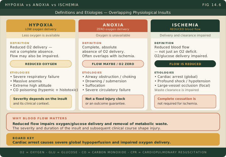
Bottom line: anoxia is absent oxygen delivery, hypoxia is reduced delivery, and ischemia is reduced blood flow, which strips the brain of oxygen and glucose at once and also stops waste clearance; cardiac arrest is a major adult cause of hypoxic-ischemic encephalopathy (HIE), with about 350,000 out-of-hospital arrests a year in the United States and roughly 10% surviving to discharge; HIE is global and produces bilateral deficits dominated by memory and cognition, while focal stroke follows an arterial territory; carbon monoxide is a double assassin that both forms carboxyhemoglobin and poisons mitochondrial cytochrome c oxidase.
The terminology drives the clinical pattern. Anoxia is complete absence of oxygen delivery, and true anoxia is uncommon: complete airway obstruction, cardiac arrest with no bystander CPR, prolonged submersion. Hypoxia is reduced but not absent delivery, as in respiratory failure, severe anemia, high-altitude exposure and CO poisoning. Because some oxygen keeps reaching the brain, hypoxic injury is usually less severe for the same duration, although prolonged severe hypoxia produces the same selective neuronal death.
Ischemia is reduced blood flow. It deprives neurons of oxygen and glucose at the same time and stops removal of metabolic waste, which is why ischemia is worse than hypoxia of comparable depth. It does not require complete cessation of flow. Cardiac arrest produces global ischemia and global oxygen failure together. Injury evolves within minutes, but no fixed number of minutes marks irreversibility in every patient.
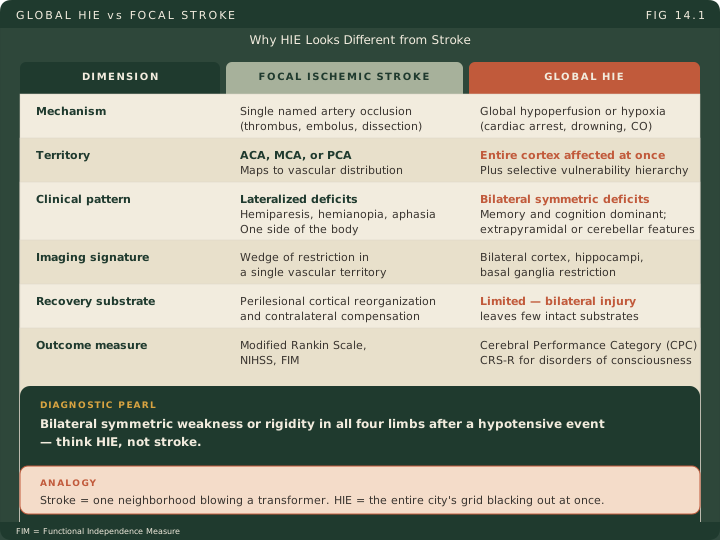
The board distinction that matters most separates HIE from stroke: global versus focal injury. Focal stroke follows occlusion of a single named artery, so the deficits map onto a vascular territory and are lateralized: hemiparesis, hemianopia, aphasia. HIE follows a global failure of perfusion or oxygenation, so the pattern is set by selective neuronal vulnerability rather than by vascular anatomy, and the deficits are bilateral and dominated by memory and cognition. The two can coexist, and bilateral infarction is possible, so neither label on its own fixes a patient’s recovery potential. Neonatal HIE means perinatal asphyxia and is a separate entity with its own treatment guidance.
Cardiac arrest is a major adult cause of HIE. About 350,000 out-of-hospital cardiac arrests occur in the United States each year, and roughly 10% of those patients survive to hospital discharge. Among survivors, the severity of neurological injury tracks the duration of impaired circulation, the quality of bystander and paramedic CPR, and the clinical course afterward. CPR circulates blood rather than restarting the heart by itself, and it can contribute to ROSC; defibrillation treats shockable rhythms and is not the final step of every arrest.
Other settings include severe hypotension, which favors watershed-zone injury because some flow is maintained, along with drowning, suffocation, strangulation, respiratory failure, opioid overdose and carbon monoxide poisoning. Strangulation compresses neck vessels as well as the airway, so it is not simply airway obstruction. Overdose can cause hypoxic injury with no documented cardiac arrest.
In drowning, the salt-water versus fresh-water distinction is clinically irrelevant to brain prognosis; submersion duration is the variable that carries prognostic weight, together with the resuscitation and the clinical course that follows. Cold-water cases can be exceptional. Cooling does lower cerebral metabolic demand, and the mammalian dive reflex shunts oxygenated blood centrally on cold-water facial immersion, but the size of that protection is not established for drowning: the human measurement comes from 37 adults during cardiac surgery, in whom cerebral metabolic rate fell with a Q10 of 2.3, roughly a 2.3-fold drop for every 10°C of cooling. Do not let water temperature alone drive a rescue or prognostic decision. ILCOR drowning recommendations, McCullough 1999
Carbon monoxide (CO) poisoning is a double assassin. CO binds hemoglobin with 200 to 250 times the affinity of oxygen, forming carboxyhemoglobin that cannot carry oxygen, and it also binds mitochondrial cytochrome c oxidase, poisoning cellular respiration directly. The dual mechanism gives CO its predilection for the globus pallidus and deep white matter. Bilateral pallidal injury is characteristic of CO poisoning but not pathognomonic: other toxic and hypoxic injuries, heroin overdose among them, produce the same lesion. Diagnose from exposure history, clinical findings and blood co-oximetry, drawn venous or arterial. Standard pulse oximetry reads carboxyhemoglobin as though it were oxyhemoglobin, so the saturation looks falsely normal. Cherry-red skin is uncommon and unreliable. The COHb concentration correlates poorly with severity and outcome, particularly once oxygen has been given, so do not use fixed COHb symptom or death bands, and do not read a low post-oxygen value as evidence of safety. Risse 1984
Give 100% oxygen to every patient with suspected CO poisoning, and consider hyperbaric treatment with expert consultation according to severity and access; selected symptomatic patients may benefit, but hyperbaric oxygen does not guarantee prevention of delayed neurological injury. CO also clears on room air, so hyperbaric treatment is not the only route of elimination. Half-lives are approximate teaching values that depend on delivery and physiology: about 4 to 6 hours on room air, 60 to 90 minutes on atmospheric 100% oxygen, and 15 to 30 minutes with hyperbaric oxygen. The measured clinical value is tighter than the textbook range: in 93 poisoned patients breathing atmospheric 100% oxygen the mean half-life was 74 minutes, range 26 to 148 minutes. Faster clearance is a kinetic endpoint and not proof of a better neurological outcome. CDC clinical guidance, Weaver 2000, ACEP 2025 policy announcement
High Yield — Definitions and Etiologies
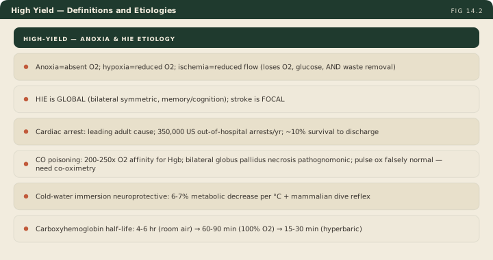
- Anoxia = absent O2; hypoxia = reduced O2; ischemia = reduced flow (loses O2 and glucose AND waste removal).
- HIE is GLOBAL: bilateral deficits, memory- and cognition-dominant. Stroke is FOCAL and follows an arterial territory.
- Cardiac arrest is a major adult cause: 350,000 US out-of-hospital arrests a year, ~10% survival to discharge.
- CO: 200 to 250x the oxygen affinity for hemoglobin, plus cytochrome c oxidase inhibition. Bilateral globus pallidus injury is characteristic but not pathognomonic.
- Pulse oximetry is falsely normal in CO poisoning; diagnose with co-oximetry on venous or arterial blood.
- COHb half-life: 4 to 6 h room air, 60 to 90 min on 100% O2, 15 to 30 min hyperbaric. Measured mean 74 min (26 to 148) on atmospheric 100% O2.
- The COHb level does not grade severity. Treat on exposure and clinical findings, not on the number.
- Drowning: submersion duration, not salt versus fresh water. Cold water lowers metabolic demand (Q10 2.3 in surgical adults) and triggers the mammalian dive reflex.
Raising the partial pressure of oxygen in the plasma to extreme levels is literally the only thing that creates enough competitive pressure to forcefully detach the carbon monoxide molecule and clear the enzymes.
— CVA-14 podcast, ~15:12
Well, because ischemia removes three vital things at once, instead of just one. Ischemia is a total cessation of blood flow.
— CVA-14 podcast, ~4:53