Before You Listen
Before You Listen
- Prerequisites: brachial plexus organization (Roots-Trunks-Divisions-Cords-Branches) and the SNAP/paraspinal/distribution rules that distinguish plexopathy from radiculopathy (covered in Episode 7); the dorsal root ganglion (DRG) principle that sensory nerve action potentials (SNAPs) are preserved in radiculopathy and abnormal in plexus or peripheral nerve lesions; nerve conduction study (NCS) basics including compound muscle action potential (CMAP) amplitude, conduction velocity (CV), and distal motor latency (DML); the difference between conduction block (focal demyelination) and reduced distal CMAP amplitude (axonal loss with Wallerian degeneration); needle electromyography (EMG) basics (fibrillation potentials, positive sharp waves, motor unit action potentials).
- Runtime: 40 minutes.
- Topic in one line: carpal tunnel syndrome (CTS) with LOAF muscles and the comparison studies (palmdiff, ringdiff, thumbdiff, second lumbrical-interossei) that detect focal median slowing the absolute latencies miss, and that outperform any single comparison when summed as the Combined Sensory Index; Bland severity (0-6); pronator and anterior interosseous nerve (AIN) syndromes; ulnar neuropathy at the elbow (UNE) with the across-elbow CV < 50 m/s rule, inching, and the dorsal ulnar cutaneous (DUC) SNAP helping distinguish UNE from Guyon canal; the Shea classification at Guyon canal; Saturday night palsy (triceps spared) and posterior interosseous nerve (PIN) syndrome (pure motor, ECRL spared); Wartenberg syndrome (pure superficial radial sensory); musculocutaneous, axillary, suprascapular (notch vs spinoglenoid), and long thoracic neuropathies; demyelinating versus axonal patterns. Memorize them cold.
Vignette. A 47-year-old right-handed woman who works as a packer presents with a four-month history of nocturnal numbness and tingling of the right thumb, index, and middle fingers. She wakes multiple times each night and shakes her hand for relief. Recently, she has dropped jars and noted thinning of the muscle at the base of her thumb. On examination, the abductor pollicis brevis is visibly atrophic, the thumb sits in the plane of the palm, and sensation is reduced over the volar surface of digits 1 through 3 but is normal over the thenar eminence itself. Phalen and Tinel signs at the wrist are positive. Nerve conduction studies show an absent median sensory nerve action potential recording from digit 2 at 14 cm, so its latency cannot be measured. The median motor distal latency is 6.8 milliseconds with a compound muscle action potential of 1.4 millivolts recording from the abductor pollicis brevis. Ulnar sensory and motor responses are normal, and the second lumbrical-interossei latency difference is 1.2 milliseconds favoring the ulnar (interossei) response. Needle electromyography of the abductor pollicis brevis reveals fibrillation potentials, positive sharp waves, and large-amplitude polyphasic motor unit action potentials with reduced recruitment.
Why is sensation preserved over the thenar eminence in this patient when the rest of the median territory is numb, what specifically does the absent median sensory response with a markedly prolonged motor distal latency and reduced compound muscle action potential tell you about severity by the Bland classification, why is the second lumbrical-interossei comparison a useful motor comparison in this scenario, and what is the most common compression site that explains a similar presentation extending up into the proximal forearm?
(Answer at the end of this chapter)
Section 1: Carpal Tunnel Syndrome — Anatomy, LOAF, and the Comparison Studies
Bottom line: CTS is the most common entrapment neuropathy; the tunnel contains the median nerve and 9 flexor tendons. LOAF identifies median-innervated hand muscles. Median–ulnar and median–radial comparisons detect subtle focal slowing that absolute latencies miss: in Robinson’s original series each single comparison found 69.7 to 75.8% of clinically affected hands and the three summed as the Combined Sensory Index found 83.1%, at 95.4 to 96.9% specificity. Sensitivity moves with protocol, cutoff and population.
The carpal tunnel is an osteofibrous canal at the wrist with the carpal bones as floor and walls and the transverse carpal ligament (flexor retinaculum) as the roof. Ten structures pass through it: the median nerve and 9 flexor tendons (4 FDS, 4 FDP, 1 FPL). The median nerve is the most superficial structure under the ligament, which is why it is the first to suffer when tunnel pressure rises. At the distal edge of the tunnel the median nerve divides into the recurrent motor branch (curving back over the thenar eminence to supply the thenar muscles) and the digital sensory branches (palmar surface of digits 1-3 and the radial half of digit 4).
The LOAF mnemonic identifies the four median-innervated muscles distal to the tunnel: Lumbricals 1-2; Opponens pollicis; Abductor pollicis brevis (APB); Flexor pollicis brevis (superficial head). The APB is the standard motor recording site because it is readily accessible and normally receives recurrent-branch innervation; median–ulnar communicating branches can alter its supply. Severe CTS with thenar atrophy produces the ape hand deformity with the thumb falling into the plane of the palm. One critical anatomic detail separates CTS from proximal median lesions: the palmar cutaneous branch arises proximal to the tunnel and runs superficial to the transverse carpal ligament rather than through the tunnel. Sensation over the thenar eminence is spared in isolated CTS. Thenar-pad numbness prompts evaluation of the palmar cutaneous branch or a proximal median lesion, including pronator syndrome; it does not exclude coexisting CTS. Lumbricals 1-2 receive motor branches from the palmar digital nerves, not the recurrent thenar branch.
Clinical Pearl — Thenar-eminence sensation is the proximal-vs-distal median tell
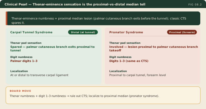
Median digit-1-to-3 numbness with a spared thenar pad is the classic CTS pattern. The palmar cutaneous branch leaves proximal to the tunnel and travels superficial to the transverse carpal ligament. Thenar-pad numbness warrants evaluation for proximal median or palmar cutaneous neuropathy, but CTS can coexist; use the whole clinical and EDX pattern.
The AANEM CTS protocol begins with median sensory and motor studies and appropriate adjacent-nerve controls. Comparative or short-segment studies are particularly useful when the initial longer-segment sensory study is normal despite clinical suspicion. Needle EMG of APB and selected C5-T1 muscles is an option, guided by the need to assess axon loss or a proximal differential, rather than mandatory in every CTS evaluation. Sensory slowing precedes motor latency abnormalities; this is not explained by smaller sensory fibers being selectively vulnerable.
Absolute latency cutoffs used in this chapter: median sensory distal latency > 3.5 ms at 14 cm, motor onset latency > 4.2 ms to APB at 8 cm, and sensory CV < 50 m/s. Interpret them against laboratory norms using the same distance, temperature and sensory onset-versus-peak convention. Note that the standard reference set in EDX-03 lists median motor onset latency up to 4.4 ms and does not state the distance it was measured over, while this chapter’s 4.2 ms is measured at a standardized 8 cm. Those are two conventions over two distances, not a contradiction; on an examination, answer with the convention the question hands you.
Comparison studies use adjacent nerves as internal controls. Palmdiff compares median–ulnar palmar mixed responses over matched distances (8 cm); an example abnormal difference is > 0.3-0.4 ms, with the median slower. Ringdiff compares median–ulnar digit-4 responses (14 cm); thumbdiff compares median–radial digit-1 responses (10 cm); example differences are > 0.4-0.5 ms. Use each laboratory’s validated technique and cutoff. In Robinson’s original series of 66 clinically affected hands and 65 control hands, each of these single comparisons detected 69.7 to 75.8% of affected hands at 95.4 to 96.9% specificity. Those figures belong to that study’s technique and cutoffs, and no one comparison has been established as the most sensitive.
The second lumbrical–interossei (2L-INT) motor comparison is especially useful in severe CTS when sensory responses or the APB CMAP are absent. Record at a common hand location and match median and ulnar stimulation distances; an example abnormal median delay is > 0.4 ms. A common recording site does not eliminate distance or temperature effects. An absent digit-2 SNAP does not necessarily make every other sensory comparison unobtainable.
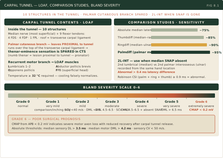
The Combined Sensory Index (Robinson CSI) sums palmdiff + ringdiff + thumbdiff, measured over 8, 14 and 10 cm respectively, to support diagnosis; it is not a severity grade. Summing the three reached 83.1% sensitivity in the original series, above any of them alone, and that is the reason to add them rather than pick a favorite. The chapter’s ≥ 0.9 ms convention requires matching the laboratory protocol (some use ≥1.0 ms).
The Bland scale grades severity from 0 to 6: 0, normal; 1, very mild, abnormalities only on sensitive comparison/segmental tests; 2, mild, sensory CV <40 m/s with normal motor latency; 3, moderate, preserved sensory response with prolonged motor latency <6.5 ms; 4, severe, absent sensory response with motor latency <6.5 ms; 5, very severe, motor latency >6.5 ms; 6, extremely severe, surface APB CMAP < 0.2 mV. Normal motor limits depend on the recording method. Severe axon loss limits thenar motor recovery, but substantial symptom improvement after release remains possible even with unrecordable responses.
Needle EMG of APB assesses motor axon loss: fibrillations and positive sharp waves indicate active denervation; long-duration motor units with reduced recruitment support chronic neurogenic change. Sampling additional muscles can help evaluate proximal lesions. Maintain hand temperature ≥32°C and use technique-matched reference values. Cooling prolongs latencies and can alter comparison differences unpredictably; it does not necessarily narrow them. Warm a 28°C hand and repeat technically inadequate studies before interpreting them.
High Yield — CTS
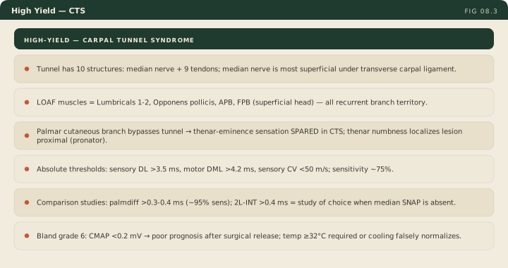
- Anatomy: 10 structures (median + 9 tendons); transverse carpal ligament roof; median nerve most superficial.
- LOAF: Lumbricals 1-2, Opponens pollicis, APB, FPB superficial head (thenar muscles via recurrent branch; lumbricals via palmar digital branches).
- Palmar cutaneous branch: classic CTS spares the thenar pad; numbness there suggests additional proximal/palmar-cutaneous involvement and does not exclude CTS.
- Absolute thresholds: 3.5 ms sensory at 14 cm, 4.2 ms motor at 8 cm and sensory CV <50 m/s are illustrative conventions; match laboratory norms, temperature and onset/peak definitions.
- Comparison studies: palmdiff >0.3-0.4 ms; ringdiff/thumbdiff >0.4-0.5 ms; 2L-INT >0.4 ms can help when sensory responses are absent. Robinson’s original series: each single comparison 69.7-75.8% sensitive, the summed CSI 83.1%, specificity 95.4-96.9%. Apply the laboratory’s own protocol.
- Bland grade 6 = APB CMAP <0.2 mV; motor recovery may be limited, while symptoms can still improve after release.
- Temperature ≥32°C: cooling can distort absolute and comparative results; warm and repeat.
The structural anatomy is absolute. If the thenar eminence is numb, the lesion must have caught the palmar cutaneous branch. Because that branch never enters the carpal tunnel, the site of compression must be proximal to the tunnel.
— EDX-08 podcast, ~6:27
Correction to the quoted teaching: thenar-pad numbness prompts additional localization; it does not exclude coexisting CTS. Localize with the full study before attributing the sensory loss to a single site.