Before You Listen
Before You Listen
- Prerequisites: lumbosacral plexus organization (lumbar plexus L1-L4 within psoas; sacral plexus L4-S4 on piriformis, with the sciatic contribution L4-S3) from Episode 7; the dorsal root ganglion (DRG) principle that sensory nerve action potentials (SNAPs) stay normal in preganglionic radiculopathy and fall in postganglionic plexus or peripheral nerve lesions; basics of nerve conduction studies (NCS) including compound muscle action potential (CMAP), sensory nerve action potential (SNAP), and the difference between conduction block (focal demyelination) and reduced distal CMAP amplitude (axonal loss with Wallerian degeneration); basics of needle electromyography (EMG) including fibrillation potentials, positive sharp waves, and the localization power of paraspinal muscle sampling; the upper-extremity entrapment patterns of Episode 8.
- Runtime: 22 minutes.
- Topic in one line: the foot drop algorithm built on the short head of the biceps femoris (spared at the fibular head, abnormal in sciatic neuropathy), the tibialis posterior (abnormal in L5 radiculopathy, normal in fibular neuropathy), and the superficial fibular SNAP (normal in L5 radiculopathy, reduced or absent in fibular or sciatic neuropathy); fibular neuropathy at the fibular head with conduction block and the accessory deep peroneal nerve variant; sciatic neuropathy with preferential fibular-division involvement (most common iatrogenic cause: total hip arthroplasty); piriformis syndrome as a diagnosis of exclusion; tarsal tunnel syndrome and its technical limitations; meralgia paresthetica (pure sensory lateral femoral cutaneous entrapment); axonal vs demyelinating prognosis. Memorize them cold.
Vignette. A 62-year-old woman who underwent right total hip arthroplasty six weeks ago presents with right foot drop and numbness over the dorsum of her right foot. On examination, ankle dorsiflexion and great toe extension are 1/5, foot eversion is 2/5, and ankle inversion and plantarflexion are full strength. Sensation is reduced over the dorsum of the foot and the lateral lower leg in the superficial fibular distribution. The right hip and gluteal muscles appear normal in bulk and strength. Nerve conduction studies show a reduced right fibular compound muscle action potential amplitude recording from the extensor digitorum brevis, with no further amplitude drop between below-fibular-head and above-fibular-head stimulation. The right tibial motor study recording from the abductor hallucis is normal in amplitude and latency. The right superficial fibular sensory nerve action potential is absent. The right sural sensory nerve action potential is normal. Needle electromyography reveals fibrillation potentials and positive sharp waves in the right tibialis anterior, peroneus longus, extensor hallucis longus, and the right short head of the biceps femoris. The right tibialis posterior, medial gastrocnemius, gluteus medius, and lumbar paraspinal muscles are all normal.
What does the abnormal short head of the biceps femoris on needle electromyography tell you about the anatomic level of the lesion, why does the absent superficial fibular sensory nerve action potential place the lesion distal to the dorsal root ganglion, what is the clinical significance of the normal tibialis posterior in this scenario, and how does the timing six weeks after total hip arthroplasty fit the most common iatrogenic mechanism for this lesion?
(Answer at the end of this chapter)
Section 1: The Foot Drop Algorithm and the Short Head of the Biceps Femoris
Bottom line: three findings resolve the foot drop differential. The short head of the biceps femoris is the only thigh muscle innervated by the fibular division of the sciatic nerve: normal in fibular neuropathy at the fibular head, abnormal in sciatic neuropathy. The tibialis posterior carries L5 through the tibial nerve: abnormal in L5 radiculopathy, normal in fibular neuropathy. The superficial fibular SNAP is normal in L5 radiculopathy (preganglionic) and reduced or absent in fibular or sciatic neuropathy (postganglionic). Read the three together, because a partial lesion can spare any one of them.
Foot drop is the central localization problem of lower-extremity electrodiagnosis. When a patient presents with ankle dorsiflexion weakness, the electromyographer distinguishes three answers: common fibular neuropathy at the fibular head, sciatic neuropathy preferentially affecting the fibular division, and L5 radiculopathy. Plexopathy, more distal lesions, and central or muscular causes belong in the differential when the examination points there. No single finding makes the diagnosis; the constellation does.
The short head of the biceps femoris is the muscle that resolves the level. It is the only muscle in the thigh innervated by the fibular division of the sciatic nerve (L5, S1). All other hamstring muscles (long head of biceps femoris, semimembranosus, semitendinosus) are innervated by the tibial division. In fibular neuropathy at the fibular head, the short head of the biceps femoris is normal because the fibular nerve is injured distal to where this muscle receives its innervation. In sciatic neuropathy with fibular predominance, the short head is abnormal because the fibular division is injured proximal to the branching point. In L5 radiculopathy, the short head is abnormal in some patients and normal in others. Learn the muscle as normal = fibular at the knee; abnormal = sciatic or radiculopathy. The abnormal result is the strong one: an isolated fibular-head lesion cannot produce it. A normal short head is supportive rather than decisive, because a partial sciatic injury, or one distal to this branch, spares the muscle, so read it with the nerve conduction studies and the rest of the needle exam.
The tibialis posterior separates L5 radiculopathy from fibular neuropathy at the fibular head. It is innervated by the tibial nerve but receives its L5 root supply, so it samples L5 outside common fibular territory. In L5 radiculopathy the tibialis posterior is abnormal because it shares the L5 root with the fibular muscles even though a different peripheral nerve supplies it. In fibular neuropathy at the fibular head the tibialis posterior is normal because the tibial nerve is unaffected. Weak ankle inversion with tibialis posterior denervation places the lesion at the root or sciatic level. A normal tibialis posterior does not send you home: the L5 root also reaches fibular-division muscles, and a partial lesion, an early study, or limited sampling can leave this muscle clean, so keep sampling other L5 muscles.
Clinical Pearl — Two muscles and a sensory response
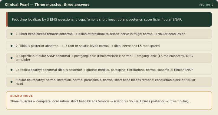
The foot drop algorithm collapses to three questions: two needle muscles and one sensory response. (1) Is the short head of the biceps femoris abnormal? If yes, the lesion is at or proximal to the sciatic nerve in the thigh. (2) Is the tibialis posterior abnormal? If yes, the lesion is at the L5 root or sciatic level, outside common fibular territory. (3) Is the superficial fibular SNAP abnormal? If yes, the lesion is postganglionic (fibular or sciatic); if it is normal, the lesion is preganglionic (L5 radiculopathy, by the DRG principle). Each abnormal answer moves the lesion. Each normal answer only narrows it, so build the diagnosis from the positive findings and treat the normal ones as supporting evidence.
Additional muscles confirm the localization. In L5 radiculopathy denervation extends beyond fibular muscles to other L5-innervated muscles supplied by different peripheral nerves. The gluteus medius and tensor fasciae latae are innervated by the superior gluteal nerve from L4-S1. Fibrillations in tibialis anterior, peroneus longus, tibialis posterior, and gluteus medius point strongly to an L5 root lesion because multiple peripheral nerves are involved but all share the L5 root. The medial gastrocnemius is primarily S1, so it is normal in L5 radiculopathy and abnormal in sciatic neuropathy that involves the tibial division.
Paraspinal muscle examination adds a fourth data point. In L5 radiculopathy, fibrillations appear in the lumbar paraspinals at the L5 level because the posterior primary ramus branches proximal to the plexus and peripheral nerve. Paraspinal fibrillations put the lesion at the root. Fibular and sciatic lesions sit distal to the posterior primary ramus and cannot themselves denervate paraspinals. Normal paraspinals, however, do not clear the root, and prior lumbar surgery makes abnormal paraspinals uninterpretable, so do not stop the study at this muscle.
The superficial fibular SNAP is the final piece. In L5 radiculopathy it is normal, because the lesion is preganglionic and the sensory axon stays attached to its cell body in the DRG (the DRG principle). In fibular and sciatic neuropathy the superficial fibular SNAP is reduced or absent, because the lesion is postganglionic and the sensory axons undergo Wallerian degeneration. Two exceptions are worth holding: a far-lateral foraminal lesion can reach the DRG itself and lower the response, and a fibular lesion that spares sensory fascicles can leave it intact. Confirm the study was technically adequate before you act on an absent response.
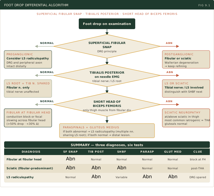
The summary algorithm: L5 radiculopathy = multiple L5-innervated muscles abnormal across different nerves (tibialis anterior, peroneus longus, tibialis posterior, gluteus medius), paraspinal fibrillations, normal superficial fibular SNAP. Fibular neuropathy at the fibular head = normal inversion, normal paraspinals, normal short head of biceps femoris, conduction block or focal slowing across the fibular head. Sciatic neuropathy (fibular-predominant) = abnormal short head of biceps femoris, normal paraspinals, normal gluteal muscles, and tibialis posterior variable, because the tibial division can be involved. These are the full patterns; an incomplete lesion produces a partial one, so match the whole picture rather than a single normal muscle.
High Yield — The foot drop algorithm
- Three diagnoses to distinguish: fibular neuropathy at fibular head, sciatic neuropathy (fibular-predominant), L5 radiculopathy.
- Short head of the biceps femoris = only thigh muscle innervated by the fibular division of the sciatic nerve. Normal = fibular at fibular head; abnormal = sciatic or radiculopathy. The abnormal result is decisive; the normal result is supportive, because a partial or distal sciatic lesion spares it.
- Tibialis posterior (tibial nerve, L5 root) = abnormal in L5 radiculopathy; normal in fibular-predominant sciatic neuropathy and in fibular at fibular head. A normal result does not clear L5; keep sampling other L5 muscles.
- Superficial fibular SNAP = abnormal in fibular and sciatic neuropathy (postganglionic); normal in L5 radiculopathy (preganglionic, DRG principle). Foraminal/DRG lesions and sensory-sparing peripheral lesions are the exceptions.
- Paraspinal fibrillations = root-level lesion (L5); fibular and sciatic lesions cannot produce them. Normal paraspinals do not clear the root, and prior lumbar surgery makes them uninterpretable.
- Gluteus medius (superior gluteal, L4-S1) = abnormal in L5 radiculopathy; normal in sciatic neuropathy (gluteal nerves arise from plexus, not sciatic trunk) and in fibular neuropathy.
- Medial gastrocnemius = primarily S1, not L5; normal in L5 radiculopathy.
So in an L5 radiculopathy, the compression is usually preganglionic, proximal to the DRG. The structural integrity of the distal sensory axon is completely preserved. So the SNAP stays normal.
— EDX-09 podcast, ~8:33
Does a normal tibialis posterior perfectly exclude an L5 radiculopathy? No, it does not perfectly exclude it. And that is a huge trap on the boards.
— EDX-09 podcast, ~6:18
But the short head of the biceps femoris is the odd one out. It alone answers to the fibular division of the sciatic nerve.
— EDX-09 podcast, ~3:49