Before You Listen
Set the stage
- Prerequisites: basic nerve conduction study (NCS) interpretation; the meaning of the compound muscle action potential (CMAP) and the sensory nerve action potential (SNAP); how a needle electromyography (EMG) examination is performed; the difference between fibrillation potentials, positive sharp waves, and motor unit action potentials (MUAPs); familiarity with F-waves and the H-reflex as late responses; the basic anatomy of a peripheral nerve including the axon, the myelin sheath, and the node of Ranvier.
- Runtime: 1 hour 25 minutes.
- Topic in one line: the Seven Key Questions framework, the axonal vs demyelinating distinction, the uniform vs non-uniform split separating hereditary from acquired disease, GBS and its three subtypes (AIDP, AMAN, AMSAN), CIDP, CMT and HNPP, MMN vs ALS, diabetic neuropathy, CIP vs CIM, and the medication and paraproteinemic neuropathies.
Vignette. A 34-year-old previously healthy man presents to the emergency department with five days of progressive ascending weakness that began with tingling in his toes and now involves both legs and his hands. He cannot climb stairs and has fallen twice today. He had a self-limited diarrheal illness three weeks ago. On exam he has symmetric lower extremity weakness 3/5, mildly reduced grip, areflexia at the patellae and Achilles, and no sensory level. Cerebrospinal fluid (CSF) shows protein 92 mg/dL with 3 cells/microL. Forced vital capacity (FVC) is 2.0 L (declining). NCS at day 6 show normal sural SNAP amplitudes bilaterally with reduced median SNAPs, mildly slowed motor conduction velocities, and absent F-waves in the tibial and median nerves.
What syndrome does this picture suggest, how should its EDX subtype be assessed, what is the named sensory pattern, what is the most important EDX prognostic feature you should look for on the CMAPs, what infectious trigger is most likely, and what is the correct treatment (and equally important, what treatment is NOT effective)?
(Answer at the end of this chapter)
Section 1: The Seven Key Questions and the Axonal-Demyelinating Split
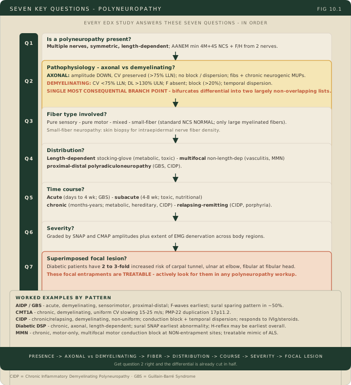
Bottom line: the Seven Key Questions organize polyneuropathy assessment: is it a polyneuropathy, axonal or demyelinating, which fiber types, what distribution, what time course, how severe, and is there a superimposed focal lesion. A demyelinating pattern is distal latency >130% ULN or CV <75% LLN; an axonal pattern loses amplitude with latency and velocity inside those limits; both together is mixed. That pair sorts a pattern and organizes the differential; diagnosing an inflammatory demyelinating polyradiculoneuropathy takes the stricter 50%/30% set in Section 2.
The Seven Key Questions organize the evaluation rather than impose a universal testing sequence. Establish whether multiple peripheral nerves are affected, then characterize the distribution: distal symmetric, multifocal, proximal-distal, or another pattern. AANEM describes a possible workup of four motor and four sensory studies across both legs and one arm, with late responses as indicated; this is not a mandatory minimum. Select and extend studies according to the examination, differential diagnosis and initial results. Sural sensory testing is useful in distal symmetric neuropathy, but the earliest detectable abnormality depends on the disease, nerve and recording technique.
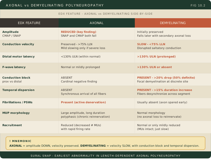
Question two asks whether the dominant physiological pattern is axonal or demyelinating. Axonal loss lowers CMAP and/or SNAP amplitudes, with relatively preserved or mildly slowed conduction in surviving fibers. The distribution need not always be length-dependent, and sensory-only or motor-only disease need not reduce both response types. Active denervation and chronic reinnervation may produce fibrillations and large, long-duration MUAPs, depending on severity and timing. Mnemonic: amplitude down, velocity relatively preserved. A low response alone does not prove irreversible axon loss; nodal conduction failure can also lower it.
A demyelinating pattern can include marked slowing, prolonged distal or F-wave latencies, conduction block and temporal dispersion. The pattern rule is distal latency above 130% of the upper limit of normal, or conduction velocity below 75% of the lower limit of normal. Meeting either one makes the study demyelinating in pattern; an abnormal study that meets neither, with reduced amplitudes, is axonal; reduced amplitudes together with a demyelinating threshold is mixed. Those two thresholds sort a pattern, and they carry no disease label with them. Diagnosing an inflammatory demyelinating polyradiculoneuropathy takes a stricter set, deliberately set further from normal because it gates immunotherapy: distal motor latency at least 50% above the ULN and motor conduction velocity at least 30% below the LLN, in more than one nerve, plus the block, dispersion and F-wave criteria of the corrected 2021 EAN/PNS framework set out in Section 2. Two threshold sets for two different jobs, and the stem will tell you which it is asking: describing a pattern, or diagnosing a disease. Conduction block has a definition, and it is segment-specific. In the AAEM consensus, definite partial conduction block requires a duration increase of no more than 30% together with either a greater than 50% drop in negative-peak amplitude or a greater than 40% drop in area, measured across the listed median, ulnar and fibular-head segments. The fibular and tibial leg segments demand more: a greater than 60% amplitude drop or a greater than 50% area drop. The tight duration cap is the point of the criterion, because it excludes temporal dispersion as the explanation for the smaller proximal response. A duration cap limits dispersion but does not by itself prove the mechanism of a reduced response. Sensory amplitudes can fall through dispersion or secondary axonal loss; preserved sensory amplitudes are not required early in every demyelinating neuropathy.
Questions three through seven sharpen the diagnosis. Question three asks which fiber types are involved: pure sensory, pure motor, mixed sensorimotor, or small-fiber (routine NCS can be normal in pure small-fiber disease because they assess large fibers; coexisting large-fiber disease may alter them). Question four asks distribution: length-dependent stocking-glove for metabolic and toxic causes; non-length-dependent multifocal for inflammatory, vasculitic, or infiltrative disease; proximal-distal polyradiculoneuropathy for GBS and CIDP. Question five asks time course: acute (days to 4 weeks; GBS), subacute (4-8 weeks; toxic, nutritional), chronic (months to years; metabolic, hereditary, CIDP), or relapsing-remitting (CIDP, porphyria). Question six grades severity by SNAP and CMAP amplitudes and EMG denervation. Question seven asks for a superimposed focal neuropathy: patients with diabetes may also have carpal tunnel syndrome, ulnar neuropathy at the elbow or fibular neuropathy at the fibular head; assess focal findings in the context of diffuse disease.
High Yield — Seven Questions and axonal-vs-demyelinating
- Seven Key Questions: presence, axonal vs demyelinating, fiber type, distribution, time course, severity, superimposed focal lesion.
- AANEM testing: four motor and four sensory studies across both legs and one arm, with late responses as indicated; extend according to the phenotype.
- Axonal pattern: low CMAP and/or SNAP amplitudes with relatively preserved conduction; denervation/reinnervation depends on timing and severity. Nodal failure can also lower amplitudes.
- Demyelinating pattern: distal latency >130% ULN, or CV <75% LLN, with marked slowing, block and/or dispersion reinforcing it; both patterns present is mixed. That pair sorts a pattern. Diagnosing an inflammatory demyelinating polyradiculoneuropathy is stricter: distal motor latency ≥50% above ULN and CV ≥30% below LLN in more than one nerve.
- AAEM definite partial conduction block: duration increase no more than 30% plus a >50% amplitude or >40% area drop in the listed median, ulnar and fibular-head segments; >60% amplitude or >50% area in the fibular and tibial leg segments.
- Mnemonic: axonal = amplitude down, velocity preserved; demyelinating = velocity slow, with block and dispersion.
- Sural SNAP: useful in distal symmetric neuropathy; not invariably the earliest abnormal test.
- DANG THERAPIST mnemonic for axonal polyneuropathy: Diabetes, Alcohol, Nutritional, GBS axonal, Toxins, Hereditary, Endocrine, Renal, Amyloid, Paraproteinemic, Infectious, Systemic, Treatment-related.
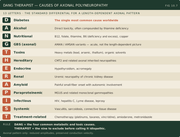
So your absolute hallmark for an axonal loss polyneuropathy is amplitude down, velocity preserved.
— EDX-10 podcast, ~7:41
Clinical qualification: This is a useful predominant pattern. Low amplitudes can also arise from conduction failure, and phenotype, timing and technique must be considered.