MSK · EP 03 · SHOULDER
Before You Listen
Episode Setup
- Topic in one line: the bony and neural pathology that closes the shoulder unit. Clavicle fractures (middle third dominant, conservative for most, surgical for open or skin-tenting); proximal humerus fractures with the Neer four-part classification (counting displaced segments by 45-degree-or-1-cm thresholds, with surgical neck most common and anatomic neck driving avascular necrosis [AVN] risk); the medial-versus-lateral scapular winging dichotomy (serratus anterior with long thoracic nerve [C5-C7] versus trapezius with spinal accessory cranial nerve [CN] XI); axillary nerve injury and the regimental badge; suprascapular nerve entrapment at the suprascapular versus the spinoglenoid notch; and thoracic outlet syndrome (TOS) with the Adson, Wright, and Roos provocative tests.
- Prerequisites: Chapter 1 (shoulder innervation and the posterior cord) and Chapter 2 (rotator cuff anatomy).
- Runtime: 1 hour.
Vignette. A 32-year-old woman undergoes excisional biopsy of a lymph node in the right posterior cervical triangle for evaluation of an enlarged node found on routine examination. Two weeks later she returns with right shoulder weakness and asymmetry. On inspection, the right scapula sits lower and farther from the spine than the left, and the inferior angle has rotated laterally toward the axilla. She cannot perform a full shoulder shrug on the right against resistance, although she can push against a wall without dramatic medial-border displacement. Sensation in the lateral deltoid (regimental badge) is preserved.
Which type of scapular winging is this, which muscle has lost innervation, which nerve is injured, why is the wall push-up relatively unrevealing in this patient, and what mnemonic captures the alternative form of winging that the boards constantly contrast with this presentation?
(Answer at the end of this chapter)
Section 1: Deltoid Anatomy and Scapular Winging — Medial Versus Lateral
Bottom line: the deltoid is a three-segment muscle (anterior driving acceleration in throwing, middle driving abduction, posterior driving deceleration), all innervated by the axillary nerve from the posterior cord at C5-C6; scapular winging is divided cleanly into medial winging (serratus anterior weakness from long thoracic nerve [C5-C7] injury, provoked by a wall push-up, captured by the MSLT mnemonic) and lateral winging (trapezius weakness from spinal accessory nerve [cranial nerve (CN) XI] injury, provoked by a resisted shoulder shrug, classically following a posterior-cervical-triangle lymph node biopsy).
The deltoid is a triangular three-segment muscle. The anterior deltoid drives the acceleration phase of throwing, firing concentrically from late cocking through ball release to bring the arm forward; strain presents as sharp anterior shoulder pain at ball release. The middle deltoid drives abduction. The posterior deltoid is the workhorse of deceleration and follow-through, contracting eccentrically to slow the arm after release; eccentric contractions generate higher force per motor unit and predispose to strain at the myotendinous junction. The entire deltoid is innervated by the axillary nerve from the posterior cord (C5-C6), which wraps the surgical neck of the humerus in the quadrilateral space and is vulnerable to stretch in fractures and dislocations.
Scapular winging is divided into medial and lateral types; the boards draw a hard line between them. The scapula is normally held flat against the posterior thoracic wall by several muscles; when the nerve supply to one fails, the scapula loses stabilization and a border lifts off the rib cage.
In medial winging, the medial border (vertical edge closest to the spine) lifts off the thoracic wall, and the scapula drifts medially and superiorly, appearing elevated and retracted. The failed muscle is the serratus anterior, which originates on the upper 8-9 ribs and inserts along the entire medial border and inferior angle of the scapula. Its primary action is protraction (pulling the scapula forward around the rib cage), and it stabilizes the medial border flat against the thorax. With paralysis, the unopposed trapezius and rhomboids pull the scapula upward and medially, and the medial border lifts off because nothing is holding it down.
The nerve is the long thoracic nerve (C5, C6, C7; “C5, C6, C7, raise your arms to heaven”; the serratus anterior is critical for overhead arm elevation). It takes a long, exposed course down the lateral chest wall, superficial to the serratus anterior, making it susceptible to traction and compression. Classic mechanisms: heavy bench pressing (bench press palsy), backpack or rucksack-strap compression against the chest wall, and iatrogenic injury during mastectomy with axillary lymph node dissection.
The gold-standard provocative maneuver is the wall push-up: the patient stands facing a wall and pushes against it with both hands. This demands aggressive serratus anterior firing to stabilize the scapula against the thorax; a paralyzed serratus immediately pops the medial border off the chest. The mnemonic MSLT consolidates the four elements: Medial winging, Serratus anterior weakness, Long Thoracic nerve injury.
Lateral winging is a different pathology with a different mechanism. The scapula is depressed and protracted, sliding inferiorly and forward around the thorax toward the axilla, with the inferior angle rotating laterally (rotary lateral winging). The visual is the opposite of medial winging: instead of the inner edge poking toward the midline, the bottom tip swings outward. The failed muscle is the trapezius, a diamond-shaped muscle with upper, middle, and lower fiber groups that elevate and retract the scapula. With paralysis, gravity and arm weight drag the scapula downward and forward.
The nerve is the spinal accessory nerve (cranial nerve XI), which runs superficially through the posterior cervical triangle just beneath skin and subcutaneous tissue. This vulnerability makes it susceptible to iatrogenic transection during procedures in the triangle. The classic vignette is a patient who undergoes a posterior cervical triangle lymph node biopsy and presents two weeks later with an inability to shrug the shoulder and a scapula drifted inferolaterally. Any neck procedure followed by shoulder dysfunction with lateral winging = spinal accessory nerve injury.
The provocative test is a resisted shoulder shrug (upper trapezius). For middle/lower trapezius, the patient squeezes the scapulae together or does a prone row; the affected scapula will not retract to midline.

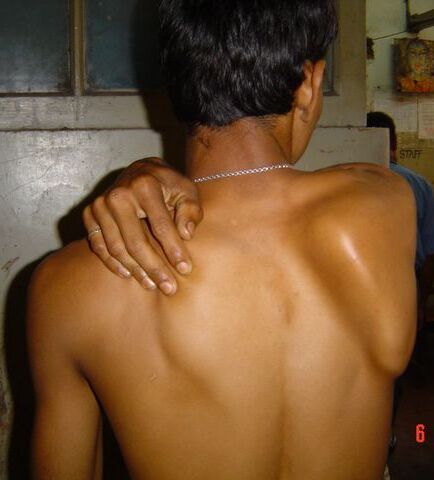
Source: Dwaipayanc, Wikimedia Commons, CC BY-SA 3.0. Clinical photograph showing medial winging of the right scapula in long thoracic nerve palsy (serratus anterior denervation).
Mnemonic — “MSLT for medial; the shrug-and-CN-XI rule for lateral”
Medial winging, Serratus anterior, Long Thoracic nerve. For lateral winging: trapezius is the “shrug muscle,” and CN XI is “the nerve you should never cut during a posterior-triangle neck biopsy.”
High Yield — Deltoid and scapular winging
- Deltoid = anterior (acceleration), middle (abduction), posterior (deceleration) — all axillary nerve, posterior cord, C5-C6.
- Medial winging = serratus anterior weakness, long thoracic nerve (C5-C7), wall push-up positive, MSLT mnemonic.
- Lateral winging = trapezius weakness, spinal accessory nerve (CN XI), resisted shrug positive, classic post-posterior-cervical-triangle biopsy.
- Long thoracic nerve mechanisms: bench press palsy, backpack strap compression, mastectomy with axillary node dissection.
- Electrodiagnostic studies (EDX) confirm injury type (neuropraxia recovers spontaneously; neurotmesis requires nerve transfer).
Lateral winging involves a completely different muscle, a completely different cranial nerve, and requires a completely different provocative test to diagnose. So mixing up the two is one of the most common and frankly most preventable errors you can make on test day.
— MSK-03 podcast, ~00:57