Before You Listen
Episode Setup
- Topic in one line: the four median nerve compression sites in pronator syndrome, the cubital tunnel as the second most common peripheral nerve entrapment, the spiral groove as the locus of humeral-shaft radial nerve injury, the Mason classification of radial head fractures with the parallel functional test that decides operative versus non-operative management of olecranon fractures, the adult distal humerus fracture and its two patient populations, the supracondylar fracture as the most common pediatric elbow fracture and the index injury for Volkmann ischemic contracture, the Panner-versus-osteochondritis-dissecans age dichotomy, the supination-flexion reduction for nursemaid’s elbow, and the CRITOE ossification sequence that lets a trainee read a pediatric elbow radiograph without missing a medial epicondylar avulsion.
- Prerequisites: elbow anatomy from MSK-04, the medial-versus-lateral epicondyle dichotomy, the elbow dislocation patterns covered in MSK-04, the Wallerian degeneration timeline from EDX-01, and the principle that compartment syndrome diagnosis depends on pain with passive stretch rather than pulselessness.
- Runtime: 1 hour.
Vignette. A 7-year-old boy falls from monkey bars onto an outstretched hand. He is brought to the emergency department with a swollen, deformed elbow. Plain radiographs reveal a displaced supracondylar fracture of the distal humerus with posterior displacement of the distal fragment. On examination, the radial pulse is faintly palpable. The capillary refill is 3 seconds at the fingertips. He cries with passive extension of the fingers in the affected hand. His distal sensation appears intact, but he cannot make the OK sign by opposing the thumb to the index finger because of weakness of thumb interphalangeal joint flexion and index finger distal interphalangeal joint flexion.
Which fracture pattern is this in the Gartland classification, which specific peripheral nerve has been injured given the inability to make the OK sign, what is the most ominous physical finding in this clinical picture, and what does that finding mandate, given that the roughly 6-hour ischemia clock has been running since the compartment pressure rose?
(Answer at the end of this chapter)
Section 1: Median and Ulnar Nerve Entrapments at the Elbow
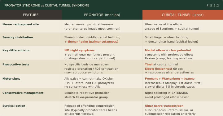
Bottom line: the median nerve at the elbow and proximal forearm passes through four sequential potential compression sites in proximal-to-distal order, namely the ligament of Struthers (an anatomical variant attaching to a supracondylar spur), the lacertus fibrosus (bicipital aponeurosis), the two heads of the pronator teres, and the fibrous arch of the flexor digitorum superficialis. Pronator syndrome shares the median sensory distribution of carpal tunnel syndrome but is distinguished by the absence of nocturnal symptoms and by sensory loss over the thenar eminence, because compression sits proximal to the palmar cutaneous branch. The anterior interosseous nerve (AIN) is a pure motor branch of the median nerve, and an isolated AIN palsy produces inability to make the OK sign because the flexor pollicis longus and the lateral half of the flexor digitorum profundus are paralyzed. Cubital tunnel syndrome is the second most common peripheral nerve entrapment in the body, behind carpal tunnel syndrome; the ulnar nerve passes through the arcade of Struthers in the medial intermuscular septum approximately 8 cm proximal to the medial epicondyle and then enters the cubital tunnel proper bounded by the medial epicondyle, the olecranon, and the arcuate ligament. The Tinel sign at the cubital tunnel and the elbow flexion test are the bedside provocative maneuvers, the Wartenberg sign (persistent small finger abduction) and Froment sign (compensatory flexor pollicis longus activation during key pinch) are the named motor signs, and management progresses from night splinting in extension through ulnar nerve transposition for refractory or motor-impaired cases.
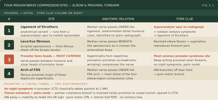
The median nerve navigates four potential compression sites as it travels from the distal arm into the proximal forearm. Memorizing the sites in proximal-to-distal order anchors the entire workup: the differential diagnosis, the imaging strategy, and the surgical target if release becomes necessary.
The first site is the ligament of Struthers, an anatomical variant present in a minority of patients. When present, the ligament connects a supracondylar spur (a small bony hook on the anteromedial distal humerus) to the medial epicondyle, with the median nerve passing underneath. Plain radiograph of the distal humerus reveals the spur. If a board stem mentions a supracondylar spur on radiograph together with median nerve symptoms, the answer is the ligament of Struthers. The second site is the lacertus fibrosus (bicipital aponeurosis), a thick fibrous sheet running off the biceps tendon beneath which the median nerve dives toward the antecubital fossa. The third site is the pronator teres muscle itself: the median nerve passes between the humeral and ulnar heads, and hypertrophy from repetitive pronation activities (screwdrivers, wringing towels, repeated forearm pronation at a manual workstation) compresses the nerve. This is the most common site of compression in pronator syndrome and the one that gives the syndrome its name. The fourth site is the arch of the flexor digitorum superficialis, where the median nerve passes under the fibrous proximal origin of that muscle.
The clinical presentation of pronator syndrome is a deep, aching pain in the proximal volar forearm with numbness in the median distribution: thumb, index finger, middle finger, and the radial half of the ring finger. The fingertip sensory map is identical to carpal tunnel syndrome, which is precisely why two specific discriminators carry the entire diagnostic logic. The first discriminator is the night symptoms pattern. Carpal tunnel syndrome classically wakes patients at 2 AM shaking their hand to chase away tingling that has built up while sleeping with wrists flexed; pronator syndrome does not produce nocturnal symptoms. The second discriminator is the palmar cutaneous branch of the median nerve, which exits the trunk proximal to the carpal tunnel and provides sensation to the thenar eminence and central palm. In carpal tunnel syndrome this branch is spared because it passes above the transverse carpal ligament rather than through the tunnel, so thenar palm sensation is preserved. In pronator syndrome the compression sits proximal to the branch point and the palmar cutaneous branch is involved, producing numbness over the thenar eminence. A stem describing numbness on the palm itself, not just the fingers, localizes the lesion proximal to the wrist and points to pronator syndrome.
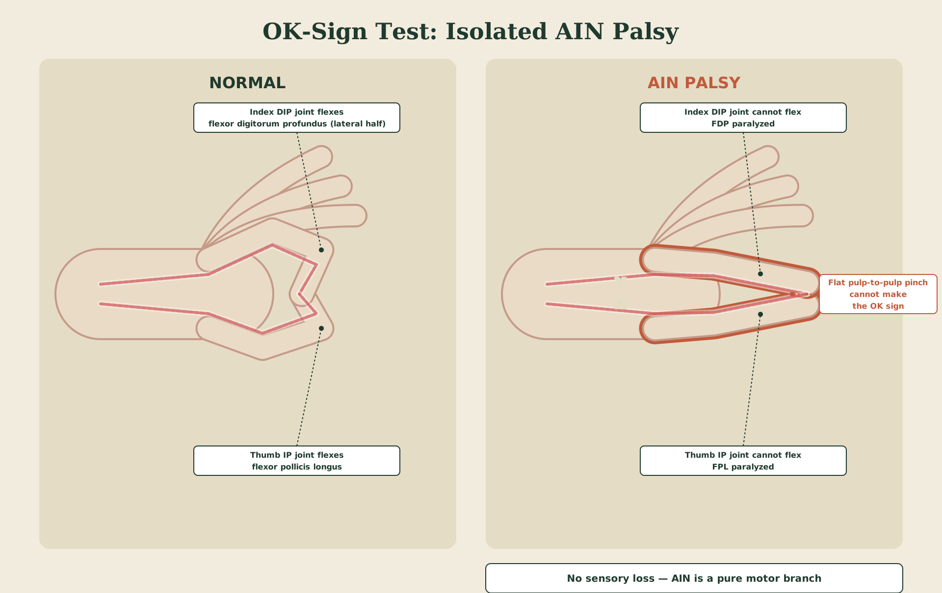
The anterior interosseous nerve (AIN) is a pure motor branch of the median nerve that arises 5 to 8 cm distal to the medial epicondyle as the median nerve runs through the cubital fossa. It supplies three muscles: the flexor pollicis longus, the lateral half of the flexor digitorum profundus (to the index and middle fingers), and the pronator quadratus. An isolated AIN palsy produces a specific bedside finding. The patient cannot make the OK sign by opposing the thumb pulp to the index finger pulp because the flexor pollicis longus cannot flex the thumb interphalangeal joint and the flexor digitorum profundus to the index finger cannot flex the distal interphalangeal joint. The thumb and index finger meet pulp-to-pulp in a pinch rather than tip-to-tip in a circle. There is no sensory loss because the AIN is purely motor. This is the pinch sign or Kiloh-Nevin sign in older textbooks. Isolated AIN palsy can be idiopathic, often a Parsonage-Turner-like neuralgic amyotrophy that resolves over many months, or due to direct compression in the proximal forearm.
Workup for proximal median nerve compression includes a plain radiograph of the distal humerus to evaluate for a supracondylar spur, and electrodiagnostic studies (electromyography and nerve conduction studies) to localize the site of compression and exclude carpal tunnel syndrome as the actual driver. Treatment is primarily conservative with activity modification, particularly elimination of the repetitive pronation that provoked symptoms. Stretching of the forearm flexors and pronator group is prescribed. If conservative management fails after a reasonable trial of at least three months, surgical release of the offending compression site is indicated.
Mnemonic — “LM, AU”
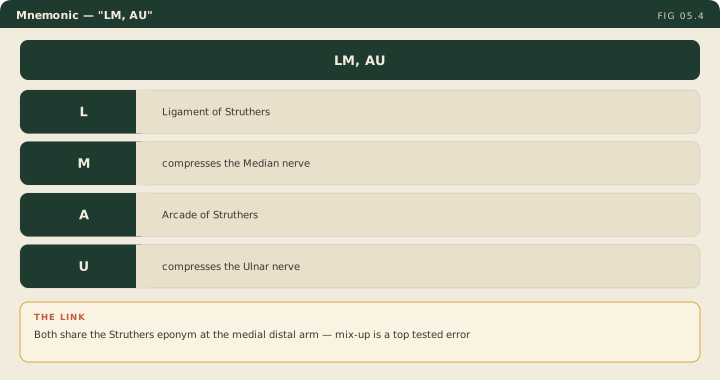
Ligament of struthers compresses the Median nerve. Arcade of struthers compresses the Ulnar nerve. The two structures share an eponym and sit in the same neighborhood at the medial distal arm; confusing them is among the most commonly tested errors on the musculoskeletal section.
Cubital tunnel syndrome is compression of the ulnar nerve at the elbow and is the second most common peripheral nerve entrapment, after carpal tunnel syndrome. Two anatomic narrowings sit in series along the medial distal arm and posterior elbow, and either one or both can drive symptoms.
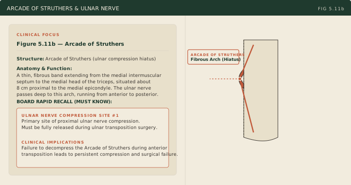
The ulnar nerve reaches the posterior elbow by passing through the arcade of Struthers, a hiatus in the medial intermuscular septum of the distal arm located approximately 8 cm proximal to the medial epicondyle. This is a different structure from the ligament of Struthers, which sits at the median nerve and connects a supracondylar spur to the medial epicondyle. The LM/AU mnemonic prevents the conflation, but the geometry helps too: the ligament hangs anteriorly across the median nerve, the arcade is a slit in the medial septum that the ulnar nerve passes through on its way posteriorly. Once posterior, the nerve enters the cubital tunnel proper, formed by the medial epicondyle, the olecranon, and the arcuate ligament that bridges between them. Compression at this level arises from nerve hypermobility with subluxation over the medial epicondyle during flexion-extension cycles, valgus stretch, osteophytes, and prolonged elbow flexion such as sleeping with elbows tucked or leaning on elbows at a desk.
Clinical presentation is medial elbow and forearm pain with numbness in the ulnar distribution: the small finger and the ulnar half of the ring finger. The ulnar nerve controls most intrinsic muscles of the hand (the interossei, the hypothenar muscles, the third and fourth lumbricals, and the adductor pollicis), and progressive neuropathy produces visible interosseous atrophy with guttering between the metacarpals when the patient lays the hand flat on a table.
Two named physical examination signs are tested repeatedly. The Froment sign tests adductor pollicis weakness. The examiner has the patient hold a piece of paper between the thumb and the lateral aspect of the index finger in a key-pinch grip and then attempts to pull the paper away. With ulnar damage the adductor pollicis is weak and cannot maintain the pinch; the brain compensates by recruiting the flexor pollicis longus (innervated by the AIN, a median branch) to flex the interphalangeal joint of the thumb into a hook that digs into the paper. Flexion of the thumb interphalangeal joint during attempted key pinch is a positive Froment sign. The Wartenberg sign is persistent small finger abduction at rest, produced when the third palmar interosseous (ulnar-innervated, normally adducts the small finger toward the ring finger) is paralyzed and the extensor digiti minimi (radial-innervated) acts unopposed.
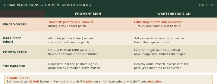
Two provocative tests anchor the bedside examination. The Tinel sign at the elbow is elicited by tapping the ulnar nerve in the cubital tunnel behind the medial epicondyle; a positive test produces electrical tingling shooting into the small finger and ulnar half of the ring finger. The elbow flexion test holds the elbow in maximum flexion for 60 seconds; reproduction of numbness or tingling in the ulnar distribution is positive. Sustained flexion raises cubital tunnel pressure and stretches the nerve over the medial epicondyle. The flexion test is analogous to the Phalen test for carpal tunnel syndrome.
Electrodiagnostic studies are the definitive workup. Nerve conduction studies typically show slowing of ulnar conduction velocity across the elbow segment, with values below 50 meters per second, or a drop of more than 10 meters per second relative to the forearm segment, considered abnormal by the AAEM practice parameter (Campbell 1999); amplitude drop and short-segment inching are adjuncts. Treatment begins conservatively. Night splinting in elbow extension or slight flexion is the cornerstone of initial management (some protocols use about 45 degrees of flexion, and the best randomized trial found no benefit of splinting over education alone: Svernlov 2009); the splint prevents the patient from sleeping with the elbow curled in deep flexion, which is the position that loads the cubital tunnel for hours at a time. Activity modification eliminates prolonged elbow flexion during the day and removes direct pressure on the medial elbow at desks, armrests, and steering wheels. If conservative management fails and there is motor atrophy or persistent symptoms, surgical intervention is indicated. The most common operation is ulnar nerve transposition, lifting the nerve out of the cubital tunnel and relocating it anteriorly under muscle, fascia, or subcutaneous tissue so that flexion no longer stretches it across the medial epicondyle.
Board answer vs. current evidence — The most common operation is ulnar nerve transposition, lifting the nerve out of the cubital tunnel and relocating it anteriorly under muscle, fascia, or subcutaneous tissue so that flexion no longer stretches it across the medial epicondyle — this is likely the board-tested answer, but more recent evidence (PMID 28953087) indicates in situ decompression and anterior transposition yield comparable outcomes, and in situ decompression has grown in popularity as an equally effective alternative. Answer the board-canonical version on the test.
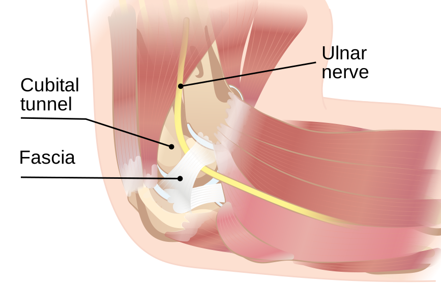
Source: InjuryMap, “Compressed ulnar nerve”, via Wikimedia Commons, CC BY-SA 4.0. https://commons.wikimedia.org/wiki/File:Compressed_ulnar_nerve.svg Medial elbow view showing the ulnar nerve coursing behind the medial epicondyle into the cubital tunnel, the site of compression in cubital tunnel syndrome.
High Yield — Pronator syndrome, AIN, and cubital tunnel syndrome
- Four median compression sites (proximal → distal): ligament of Struthers (supracondylar spur), lacertus fibrosus, pronator teres heads, arch of flexor digitorum superficialis.
- Pronator vs carpal tunnel: pronator syndrome has no night symptoms and thenar eminence numbness (palmar cutaneous branch involvement); carpal tunnel has both night symptoms and spared thenar sensation.
- AIN palsy = pure motor branch of the median nerve; cannot make OK sign (loss of flexor pollicis longus + flexor digitorum profundus to index); no sensory loss.
- Workup: radiograph for supracondylar spur + electrodiagnostic studies for site localization.
- Cubital tunnel syndrome = second most common peripheral nerve entrapment; ulnar nerve at the elbow.
- Arcade of Struthers = medial intermuscular septum hiatus approximately 8 cm proximal to the medial epicondyle (not the same as the ligament of Struthers).
- Cubital tunnel proper = bounded by medial epicondyle, olecranon, and arcuate ligament.
- Wartenberg sign = persistent small finger abduction (paralyzed third palmar interosseous, unopposed extensor digiti minimi).
- Froment sign = thumb interphalangeal flexion during key pinch (paralyzed adductor pollicis, compensatory flexor pollicis longus).
- Treatment: night splinting in extension first; ulnar nerve transposition for refractory or motor cases.
Board Trap — Calling cubital tunnel symptoms a C8 radiculopathy
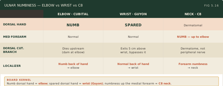
A stem describes ring and small finger paresthesias and asks for the diagnosis. Both a C8 radiculopathy and an ulnar neuropathy can produce ring and small finger numbness. The discriminator is sensation over the dorsal hand. The dorsal cutaneous branch of the ulnar nerve provides sensation to the ulnar dorsum of the hand and exits the ulnar nerve roughly 5 cm proximal to the wrist. A lesion at the elbow takes out this branch (dorsal hand numbness present); a lesion at the wrist within Guyon canal spares it (dorsal hand sensation preserved). A C8 radiculopathy produces dermatomal numbness of the entire small finger and may extend up the medial forearm, a different sensory pattern than ulnar neuropathy.
Palm numbness, not just fingertip numbness, forcefully pulls the lesion proximal to the wrist. If the palm is numb, you are not dealing with a simple carpal tunnel.
— MSK-05 podcast, ~7:19