MSK · EP 13 · FOOT
Before You Listen
Episode Setup
- Topic in one line: the high-yield foot conditions that pivot on small anatomic landmarks (medial calcaneal tubercle, third intermetatarsal space, Lisfranc keystone) and on the vascular/biomechanical map that decides whether a fracture heals or fails.
- Prerequisites: ankle and foot bony anatomy, the deep peroneal/tibial/sural innervation map, and the windlass mechanism.
- Runtime: 1 hour 21 minutes.
Vignette. A 37-year-old recreational runner doubled her weekly mileage in preparation for her first marathon. Six weeks into training she develops insidious pain over the dorsum of the midfoot, slightly medial to the second metatarsal shaft. The pain is worst in the second half of long runs and present the next morning when she steps out of bed. A plain radiograph one week after symptom onset is read as normal. Examination shows focal tenderness over the second metatarsal shaft without ecchymosis. She has irregular menstrual cycles and admits to disordered eating during her training cycle.
What is the most likely diagnosis, why was the radiograph negative, what imaging study should be ordered next, what systemic syndrome should be screened for, and what high-risk stress fracture sites must be excluded by careful clinical reasoning?
(Answer at the end of this chapter)
Section 1: Foot Architecture, Stress Fractures, and the Torg Classification
Bottom line: the foot has three arches (medial longitudinal, lateral longitudinal, transverse), the second and third metatarsals are the most commonly stressed bones because the second metatarsal is the longest and most rigidly fixed at the tarsometatarsal joint, the high-risk stress fracture locations (femoral neck tension side, anterior tibial cortex with the dreaded black line, fifth metatarsal Jones zone, navicular, talus) are the testable group, and the Torg classification splits fifth metatarsal base injuries into a benign tuberosity avulsion (zone 1) and watershed Jones/diaphyseal stress fractures (zones 2 and 3) with a high nonunion rate.
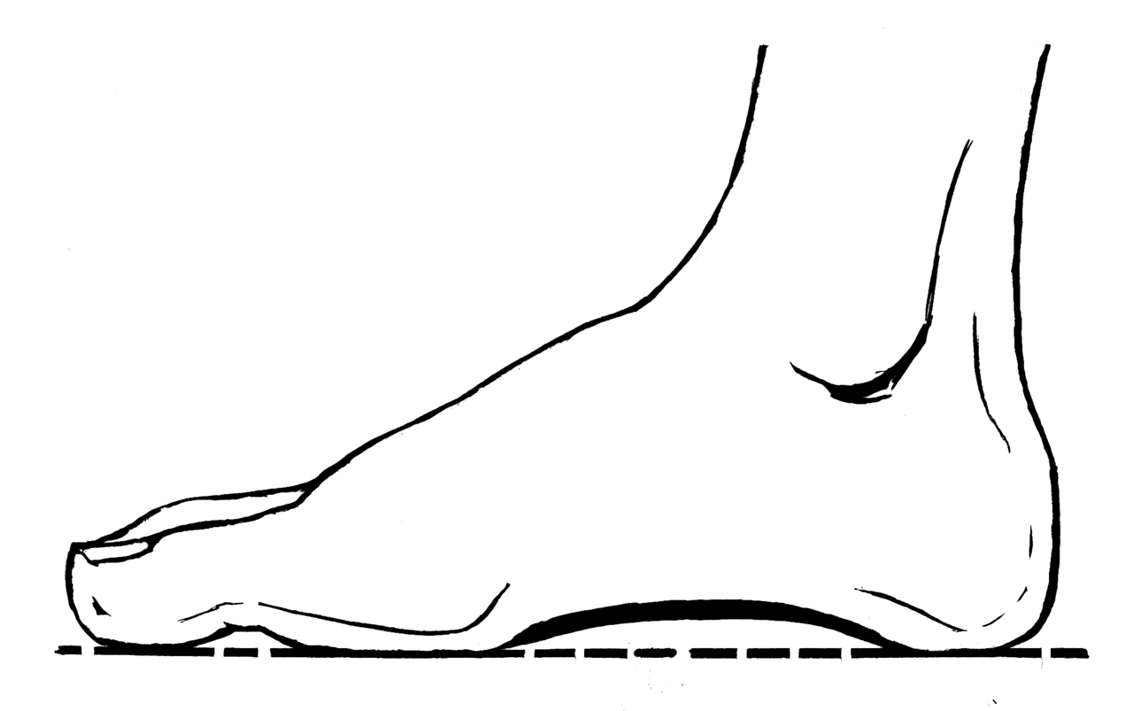
The foot has three arches: the medial longitudinal arch (calcaneus, talus, navicular, three cuneiforms, first three metatarsals), the lateral longitudinal arch (calcaneus, cuboid, fourth and fifth metatarsals), and the transverse arch (across the cuneiforms and metatarsal bases). The medial arch is the highest and the dynamic engine of gait; the transverse arch contributes to forefoot load distribution. The plantar fascia and the posterior tibial tendon are the two soft tissue structures that maintain the medial longitudinal arch.
Stress fractures are the result of repetitive submaximal loading that overwhelms the bone’s ability to remodel. Microdamage accumulates faster than osteoclastic resorption and osteoblastic repair can keep pace. The tibia is the most common location for stress fractures in the lower extremity overall because it absorbs most axial loading during ambulation and running. The second and third metatarsals are next, historically called march fractures because they were first described in military recruits subjected to forced marching. Runners who rapidly increase their weekly mileage are the modern equivalent. The second metatarsal is preferentially affected because it is the longest and most rigidly fixed at the tarsometatarsal (TMT) joint, making it the primary weight-bearing beam of the forefoot; it cannot dissipate force by moving the way the first and fifth metatarsals can.
Source: Pearson Scott Foresman archive donated to Wikimedia Foundation, Public Domain.
High-risk stress fracture locations carry a disproportionate rate of complications, including nonunion, displacement, and avascular necrosis. The femoral neck is the first high-risk location. Within the femoral neck, the critical distinction is between the tension side (superior cortex) and the compression side (inferior cortex). A tension-side fracture is dangerous because tensile forces actively pull the fracture apart with every step, creating high risk of displacement and avascular necrosis of the femoral head. Tension-side femoral neck stress fractures require immediate non-weight-bearing and often surgical fixation. Compression-side fractures heal with protected weight-bearing.
The anterior tibia is the second high-risk location. A stress fracture of the anterior tibial cortex produces the dreaded black line on lateral radiographs, a transverse lucency representing a chronic stress fracture that has failed to unite. The anterior cortex of the tibia is under tension during weight-bearing because the tibia bows slightly forward, and that tensile environment prevents healing. These fractures are notoriously resistant to conservative care and frequently require intramedullary nailing for union. The fifth metatarsal base, the navicular, and the talus round out the high-risk list. The navicular has a central watershed zone of relative hypovascularity (analogous to the scaphoid) that predisposes to nonunion. Talar stress fractures are uncommon but carry a high risk of avascular necrosis given the precarious blood supply.
Relative Energy Deficiency in Sport (RED-S, formerly the female athlete triad) is the most important systemic risk factor for stress fracture. RED-S describes inadequate caloric intake relative to exercise energy expenditure, producing hormonal disruption including menstrual dysfunction, decreased estrogen, and impaired bone mineral density. Any female athlete with a stress fracture should be screened for disordered eating, menstrual history, and bone density. Plain radiographs are frequently normal in the first 2 to 3 weeks. Magnetic resonance imaging (MRI) is the gold standard for early diagnosis because it detects bone marrow edema. Bone scintigraphy is an alternative.
The Torg classification organizes fractures at the base of the fifth metatarsal into three zones, and zone identification drives every management decision. Zone 1 is the tuberosity avulsion fracture, sometimes called the pseudo-Jones fracture. The fracture line lies within the tuberosity proximal to the fourth-fifth intermetatarsal articulation. The mechanism is an inversion ankle injury in which the peroneus brevis tendon (or the lateral band of the plantar fascia) avulses a fragment from the tuberosity. Zone 1 is by far the most common fifth metatarsal fracture; blood supply to this region is robust, healing is reliable, and treatment is conservative (hard-soled shoe or walking boot, weight-bearing as tolerated, 4 to 6 weeks). Zone 2 is the true Jones fracture, located at the metaphyseal-diaphyseal junction, typically within 1.5 cm of the tuberosity, and involves the fourth-fifth intermetatarsal articulation. This zone is a vascular watershed between metaphyseal blood supply and diaphyseal nutrient artery, producing a 15 to 30 percent nonunion rate. Conservative management with a non-weight-bearing short leg cast for 6 to 8 weeks is the standard starting point, but operative intramedullary screw fixation is frequently chosen upfront for high-demand athletes. Zone 3 is a diaphyseal stress fracture, just distal to the Jones zone, seen in distance runners and ballet dancers; it is similarly prone to delayed union and is often operative.

Board Trap — “Inversion injury plus fifth metatarsal pain equals Jones”
A vignette describes acute lateral foot pain after an inversion ankle injury with a fracture at the base of the fifth metatarsal. The trap answer is “Jones fracture, surgical fixation.” The right answer depends on the Torg zone. An avulsion at the proximal tuberosity is Zone 1 (pseudo-Jones) and is treated conservatively in a hard-soled shoe or walking boot. The true Jones fracture is Zone 2 at the metaphyseal-diaphyseal junction. Mislabeling Zone 1 as Jones leads to unnecessary surgery; the answer is to identify the zone radiographically before deciding.
High Yield — Foot architecture, stress fractures, Torg
- Three arches: medial longitudinal, lateral longitudinal, transverse.
- Tibia = most common stress fracture site overall. Second and third metatarsals = march fractures.
- High-risk locations: femoral neck tension side (superior cortex), anterior tibial cortex (dreaded black line), fifth metatarsal Jones zone, navicular, talus.
- RED-S (formerly female athlete triad) = low energy availability + menstrual dysfunction + low bone mineral density.
- MRI is the gold standard for early stress fracture diagnosis (bone marrow edema).
- Torg Zone 1 (pseudo-Jones tuberosity avulsion) heals conservatively. Zone 2 (true Jones) and Zone 3 (diaphyseal stress) sit in a vascular watershed and often need an intramedullary screw.
A stress fracture on the tension side, the superior cortex, is incredibly dangerous. It’s a ticking time bomb, because those tensile forces are actively pulling the microscopic fracture apart with every single step. Conversely, the compression side, the inferior cortex, is a much more biologically forgiving environment, because the forces are pushing the bone edges together.
— MSK-13 podcast, ~5:21