MSK · EP 20 · RHEUM
Before You Listen
Episode Setup
- Topic in one line: the three workhorse rheumatologic diseases of the joint. Rheumatoid arthritis (RA) is a symmetric autoimmune synovitis that erodes the metacarpophalangeal (MCP) and proximal interphalangeal (PIP) joints while sparing the distal interphalangeal (DIP) joint. Osteoarthritis (OA) is degenerative cartilage wear that loves the DIP joint and weight-bearing surfaces. The two crystal arthropathies (gout and pseudogout) are separated by a single sentence about needles versus rhomboids and yellow versus blue.
- Prerequisites: synovial joint anatomy, the three connective tissue layers of cartilage and capsule, basic immunology of immunoglobulin M and major histocompatibility complex class II, and synovial fluid analysis from your general medicine rotation.
- Runtime: 52 minutes 30 seconds.
Vignette. A 56-year-old woman presents with three months of bilateral hand pain and swelling. She reports stiffness on waking that takes about ninety minutes to loosen and improves once she has been moving around the house. Examination shows soft, boggy swelling at both second and third MCP joints and both wrists, with tenderness on compression of the MCP row. The DIP joints are clinically normal. Anti-cyclic citrullinated peptide (anti-CCP) antibody is positive, rheumatoid factor (RF) is positive, and erythrocyte sedimentation rate is elevated. Her primary care physician started low-dose methotrexate and folic acid two weeks ago and now refers her for inpatient elective elbow surgery requiring general anesthesia.
Which serologic finding is most specific for the underlying diagnosis, what cervical spine evaluation is required before her airway is manipulated, what is the duration of morning stiffness threshold that distinguishes her disease from osteoarthritis, and what tuberculosis screening step would be required if her physician chose to escalate her therapy to a tumor necrosis factor (TNF) inhibitor?
(Answer at the end of this chapter)
Section 1: Rheumatoid Arthritis — Pattern, Pannus, and Pre-Op Cervical Risk
Bottom line: rheumatoid arthritis is a symmetric, MCP/PIP/wrist-predominant inflammatory arthritis driven by synovial pannus that spares the DIP joint, declares itself with morning stiffness over sixty minutes, is most specifically confirmed by anti-CCP antibody, and carries a board-mandatory cervical spine question. Atlantoaxial subluxation must be excluded with flexion-extension radiographs before any patient with established RA is intubated.
Rheumatoid arthritis (RA) is a chronic, systemic, autoimmune inflammatory arthropathy whose target tissue is the synovial membrane. The single highest-yield differentiating feature is the joint distribution. RA produces a symmetric polyarthritis that preferentially attacks the metacarpophalangeal joints, the proximal interphalangeal joints, and the wrists, and characteristically spares the distal interphalangeal joints. If a stem describes symmetric swelling and pain in the MCP and PIP joints with the DIP joints completely uninvolved, the answer is rheumatoid arthritis (Figure 20.1).

Morning stiffness in RA lasts greater than 60 minutes, is worst on waking, and improves with movement. Inflammatory mediators accumulate overnight and require sustained motion to disperse. Osteoarthritis behaves the opposite way: stiffness is brief (under 30 minutes), pain worsens with activity, and rest provides relief. The 30-minute versus 60-minute duration is one of the most frequently tested distinguishing facts on the board exam.
The pathophysiology centers on pannus. The immune system attacks the synovial lining and produces a hypertrophied, inflamed tissue mass that invades and erodes articular cartilage, subchondral bone, tendons, and ligaments from the joint margins inward. Pannus is the destructive engine of RA. Pannus is what produces the four classic hand deformities the boards expect on sight (Figure 20.2). Swan-neck deformity is hyperextension at the PIP with flexion at the DIP from dorsal subluxation of the lateral bands. Boutonnière deformity is the mirror image: PIP flexion with DIP hyperextension from rupture of the central slip and volar migration of the lateral bands. Ulnar deviation (ulnar drift) at the MCP joints comes from chronic synovitis stretching the radial collateral ligaments. Z-thumb is MCP flexion with interphalangeal hyperextension. The combination of MCP ulnar deviation with swan-neck or boutonnière deformities is virtually pathognomonic for advanced RA.
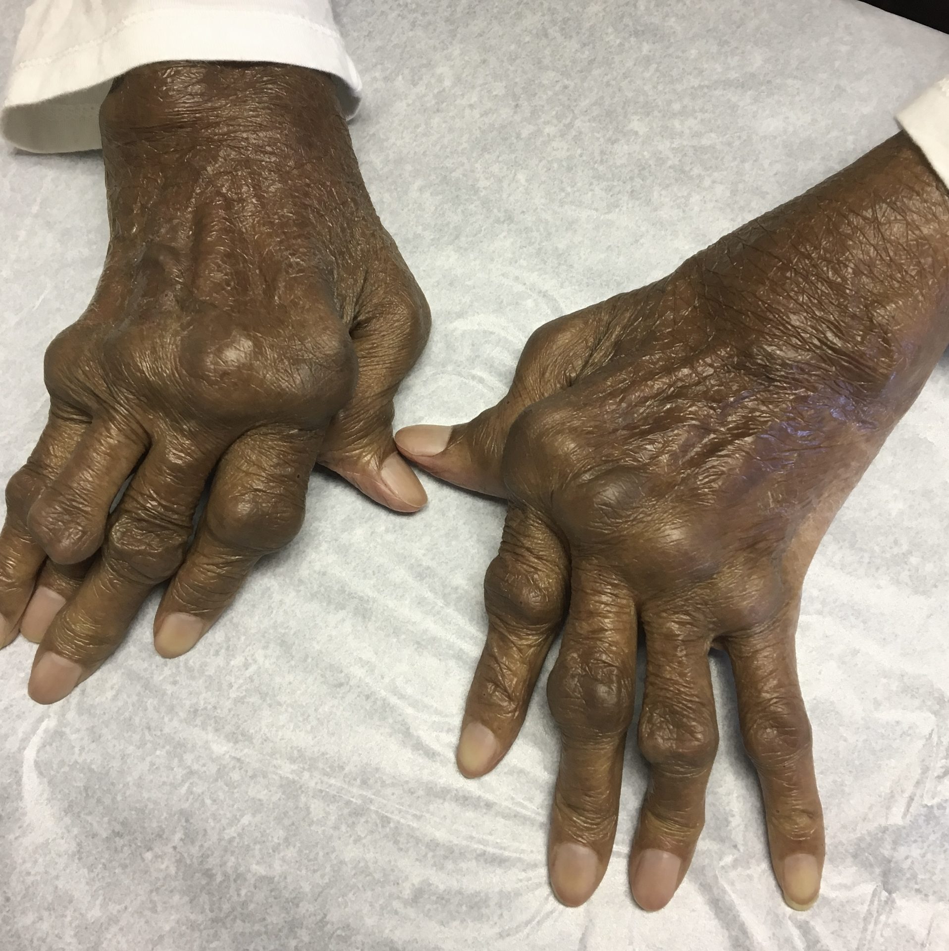
Source: Dr. Gannavappu Narasimhamurthy, Wikimedia Commons, CC0 1.0 (Public Domain Dedication)
The cervical spine question is mandatory. Atlantoaxial subluxation at C1-C2 develops when chronic synovitis weakens the transverse ligament that normally holds the odontoid against the anterior arch of C1. The atlantodental interval (ADI), the distance between the posterior surface of the anterior arch of C1 and the anterior surface of the odontoid, is the key measurement. Normal ADI is less than 3 mm. An interval greater than 3 mm indicates instability. Any patient with established RA scheduled for general anesthesia requiring intubation must have cervical spine flexion-extension radiographs before the anesthesiologist extends the neck for laryngoscopy. Missing this can produce spinal cord compression during airway management.
The serologies separate sensitivity from specificity. Rheumatoid factor (RF) is an immunoglobulin M antibody directed against the fragment crystallizable (Fc) portion of immunoglobulin G; it is present in 70-80% of RA patients but is also positive in Sjögren syndrome, systemic lupus erythematosus, hepatitis C, bacterial endocarditis, and healthy elderly individuals. Anti-cyclic citrullinated peptide (anti-CCP) antibody is the most specific serologic marker, with specificity exceeding 95%. A patient positive for both RF and anti-CCP carries the highest diagnostic certainty and a more aggressive, erosive disease course. If a board question asks the most specific test for RA, the answer is anti-CCP, every time.
RA is systemic. Extra-articular manifestations include rheumatoid nodules over pressure points (olecranon, extensor forearms), interstitial lung disease, pleural effusions, scleritis and episcleritis, accelerated atherosclerosis (a leading cause of mortality), anemia of chronic disease, and Felty syndrome (RA + splenomegaly + neutropenia). Caplan syndrome is the eponym for RA plus pneumoconiosis, classically in a coal miner with cavitating rheumatoid pulmonary nodules that mimic tuberculosis or malignancy. The piano key sign at the wrist is the bouncy distal ulna in pronation from chronic destruction of the distal radioulnar joint ligaments; surgical options when conservative care fails include the Darrach procedure (distal ulnar resection) or the Sauvé-Kapandji procedure (distal radioulnar joint fusion with ulnar pseudarthrosis).
High Yield — Rheumatoid Arthritis
- Pattern: symmetric polyarthritis of MCP, PIP, wrists; DIP spared.
- Morning stiffness >60 min, improves with activity (vs <30 min in OA, worsens with use).
- Anti-CCP = most specific (>95%); RF = sensitive but not specific.
- Atlantoaxial subluxation (ADI >3 mm) = mandatory pre-intubation flexion-extension X-rays.
- Hand deformities: swan neck (PIP up, DIP down), boutonnière (PIP down, DIP up), ulnar deviation, Z-thumb.
- Caplan syndrome = RA + pneumoconiosis with cavitating nodules.
- Piano key sign = distal radioulnar instability; treat with Darrach or Sauvé-Kapandji.
Mnemonic — DIP-spared = RA, DIP-loved = OA
If the stem describes the distal interphalangeal joints as the swollen, painful, or deformed knuckles, the disease is osteoarthritis (Heberden nodes). If the DIP joints are completely silent and the MCP and wrist take all the inflammation, the disease is rheumatoid arthritis. The boundary between these two diseases lives at the DIP joint.
If that patient has undiagnosed atlantoaxial instability, the extreme mechanical force of extending the neck during intubation can cause the weakened transverse ligament to fail completely. The atlas subluxates, the spinal cord is crushed on the operating table, and the patient wakes up paralyzed, or worse, they do not wake up at all.
— MSK-20 podcast, ~19:43