Before You Listen
Episode Setup
- Topic in one line: the two non-inflammatory pain disorders most likely to walk into PM&R clinic. Complex regional pain syndrome (CRPS) is a chronic neuropathic pain syndrome with sensory, vasomotor, sudomotor/edema, and motor/trophic features following an inciting event (most often distal radius fracture); myofascial pain syndrome is a regional muscle pain disorder driven by trigger points in taut bands. Both demand precise diagnostic criteria, both demand multimodal rehabilitation, and both have specific board-tested treatment dos and don’ts.
- Prerequisites: basic peripheral nerve anatomy, sympathetic nervous system anatomy (stellate ganglion at C7-T1 with the C6 Chassaignac tubercle as the needle landmark, lumbar sympathetic chain along L2-L4), the gate control theory of pain, the three-phase bone scan, and electrodiagnostic basics from prior MSK and EDX chapters.
- Runtime: 40 minutes.
Vignette. A 52-year-old woman sustained a left distal radius fracture six months ago after a fall. Her cast was removed at six weeks. She now presents to clinic with continuing pain in the left hand and wrist that is disproportionate to the original injury. Examination reveals allodynia to light touch, temperature asymmetry with the left hand 1.5 degrees Celsius cooler than the right, persistent edema, restricted range of motion in the wrist and fingers, glossy skin, and altered nail growth. Plain radiographs show patchy periarticular osteoporosis of the left hand. Nerve conduction studies are normal.
What diagnostic criteria does this patient meet, what is the most commonly tested imaging study to support the diagnosis, what type of CRPS does this represent and why, what is the single most important rehabilitation intervention, and what was the established prophylactic recommendation that could have reduced her risk of developing this condition after the original distal radius fracture?
(Answer at the end of this chapter)
Section 1: CRPS — Type 1 vs Type 2 and the Budapest Criteria
Bottom line: CRPS Type 1 (formerly reflex sympathetic dystrophy, RSD) occurs without confirmed nerve injury; CRPS Type 2 (formerly causalgia) occurs with confirmed peripheral nerve injury; the clinical presentation, criteria, and treatment are identical for both types; the Budapest criteria require continuing pain disproportionate to the inciting event plus symptoms in 3 of 4 categories and signs in 2 of 4 categories at evaluation, with no other better explanation.
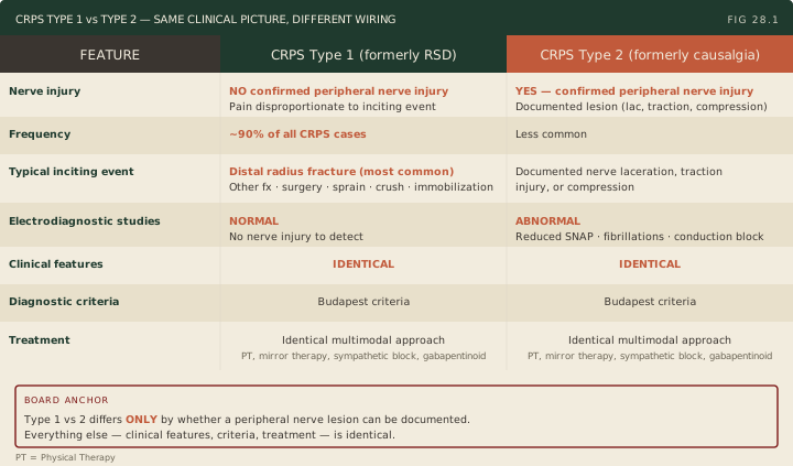
Complex regional pain syndrome (CRPS) is a chronic pain condition characterized by pain disproportionate in severity, duration, or distribution to the inciting event, accompanied by sensory, vasomotor, sudomotor, edema, and motor/trophic changes. CRPS divides into two types based on the presence or absence of confirmed nerve injury (Figure 28.1).
The most common precipitating event for CRPS is distal radius fracture. Other triggers include other fractures, surgery, sprains, crush injuries, and immobilization. CRPS most commonly affects the distal extremities and occurs in either the upper or lower extremity, though the upper extremity is affected more often. Women are affected roughly three to four times as often as men. The age of onset spans a wide range rather than one narrow peak: an American population study found a median onset in the mid-40s, while a larger Dutch population study found the highest incidence in postmenopausal women in their 60s. The pathophysiology is multimechanistic: peripheral and central sensitization, neurogenic inflammation, sympathetic nervous system dysfunction, and cortical reorganization with shrinkage of the somatosensory representation of the affected limb. Substance P and calcitonin gene-related peptide drive the neurogenic edema, vasodilation, and sweating changes.
The Budapest criteria are the international consensus diagnostic criteria and require all four conditions (Figure 28.2):
- Continuing pain disproportionate to the inciting event.
- Symptoms (patient-reported) in at least 3 of 4 categories: sensory (hyperalgesia, allodynia), vasomotor (temperature asymmetry, skin color changes), sudomotor/edema (swelling, sweating asymmetry), and motor/trophic (decreased range of motion, weakness, tremor, dystonia, hair/nail/skin changes).
- Signs (examiner-documented) in at least 2 of 4 categories at the time of evaluation, drawn from the same four categories.
- No other diagnosis that better explains the findings.
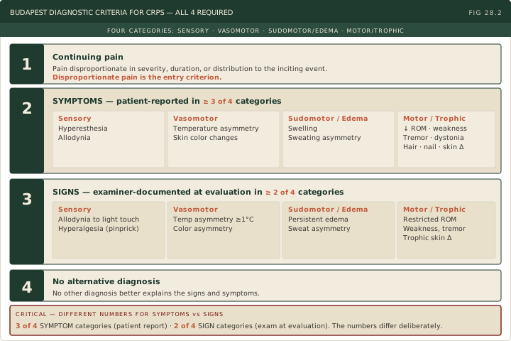
Note the distinction between criteria 2 and 3. Criterion 2 requires symptoms in 3 of 4 categories (what the patient reports). Criterion 3 requires signs in 2 of 4 categories (what the examiner observes and documents). The numbers differ deliberately.
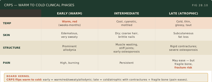
The clinical features evolve through three stages, though the modern view treats progression as variable rather than rigidly sequential. Acute (early) phase, weeks to months: warm, red, edematous, sweaty extremity with burning pain and prominent allodynia. The limb appears swollen and shiny, reflecting increased sympathetic activity and peripheral vasodilation. Intermediate (dystrophic) phase, months: limb transitions from warm to cool; skin becomes cool, cyanotic or mottled, and dry; edema persists; muscle wasting and joint stiffness develop; hair becomes coarse or is lost; nails become brittle and ridged; osteoporosis progresses. Late (atrophic) phase: skin becomes thin, glossy, and atrophic; subcutaneous tissue is lost; severe joint contractures and muscle atrophy develop; pain may paradoxically decrease somewhat as tissue atrophy progresses, but functional impairment is severe; significant osteoporosis predisposes to pathologic fracture.
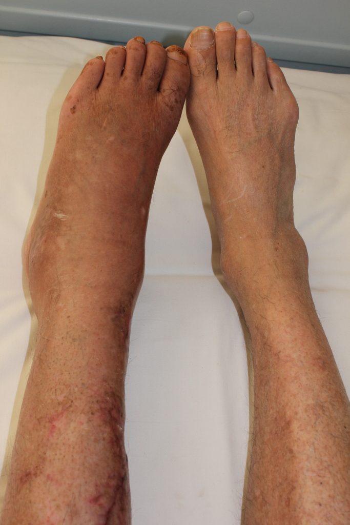
Source: Timsong311, “Complex regional pain syndrome”, via Wikimedia Commons, CC BY-SA 3.0 / GFDL. https://commons.wikimedia.org/wiki/File:Complex_regional_pain_syndrome.jpg
Many patients present with features from multiple stages simultaneously, and the average time from symptom onset to diagnosis is several months. Early recognition and treatment improve outcomes. Children and adolescents differ from adults: the lower extremity is the usual site, girls outnumber boys about 4:1, the bone scan is unreliable (often normal or showing decreased uptake), treatment is physical therapy with TENS, biofeedback and a tricyclic rather than blocks, and the prognosis is good, whereas adult-onset CRPS carries a poor prognosis. A high index of suspicion in any patient with pain and autonomic features disproportionate to the inciting event is essential.
High Yield — CRPS Classification and Diagnosis
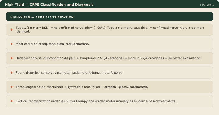
- Type 1 (formerly RSD) = NO confirmed nerve injury (~90% of cases). Type 2 (formerly causalgia) = confirmed nerve injury. Clinical features and treatment are identical.
- Distal radius fracture is the single most common precipitant.
- Budapest criteria: continuing pain disproportionate + symptoms in 3 of 4 categories + signs in 2 of 4 categories + no other better explanation.
- Categories: sensory, vasomotor, sudomotor/edema, motor/trophic.
- Three stages: acute (warm/red) → dystrophic (cool/blue) → atrophic (glossy/contracted).
- Cortical reorganization is the modern mechanistic anchor for mirror therapy and graded motor imagery as evidence-based interventions.
If you demanded three out of four for signs, you would fail patients who genuinely have the disease simply because their sudomotor or vasomotor changes or having a quiet hour while they happen to be sitting on your table. So the criteria drop the bar to two of four for the harder evidence to account for that temporal fluctuation.
— MSK-28 podcast, ~10:18
The diagnostic criteria are identical. The treatment protocols are identical. The only separator between type 1 and type 2 is the electrodiagnostic evidence of nerve damage.
— MSK-28 podcast, ~3:48