PO · EP 04 · PROSTHETICS
Before You Listen
Before You Listen
- Prerequisites: familiarity with the Medicare Functional Classification System (K0 through K4); the surgical concepts of myodesis versus myoplasty from Episode 3; the gross anatomy of the proximal tibia and fibula (tibial crest, tibial tubercle, medial tibial flare, fibular head, fibular neck); the course of the common peroneal (fibular) nerve as it wraps around the fibular neck; the eight subphases of the gait cycle from Episode 1 (initial contact, loading response, midstance, terminal stance, preswing, initial swing, midswing, terminal swing); the basic concept of the ground reaction force (GRF) vector and its relationship to a joint axis.
- Runtime: 1 hour 2 minutes.
- Topic in one line: the four interdependent elements of every transtibial prosthesis (socket, liner, suspension, foot-ankle), with the patellar tendon bearing (PTB) socket and the modern total surface bearing (TSB) socket as the two foundational designs; the pressure-tolerant areas that are loaded and the pressure-sensitive areas that are relieved (with the fibular head and the common peroneal nerve at the top of the danger list); the five elastomeric liner materials and their distinguishing mechanical properties; the eight transtibial suspension systems from supracondylar cuff to elevated vacuum (VASS); the prosthetic feet matched to K-level from SACH (K1) to energy-storing carbon fiber feet and powered ankles (K3-K4); the three stages of alignment (bench, static, dynamic); and the systematic gait deviation algorithm that distinguishes prosthetic causes from patient-related causes for the most commonly tested transtibial gait abnormalities.
Vignette. A 58-year-old man with a six-month-old right transtibial amputation from a work-related crush injury returns to clinic two weeks after receiving his definitive prosthesis. He reports a new burning numbness over the dorsum of his right foot and difficulty clearing his toe during swing phase. On examination, the residual limb is well-healed with a posterolateral surgical scar, the fibula has been cut approximately 2 cm shorter than the tibia, and the tibial crest has been beveled. Strength testing reveals new weakness of ankle dorsiflexion and toe extension on the right. The socket fits snugly with a pin-lock suspension and a silicone gel liner. There is point tenderness over the proximal lateral leg just distal to the fibular head when the socket is removed.
Which specific anatomic structure is being compressed by the prosthesis, what is the named clinical syndrome that this presentation represents, what is the single most important socket modification required, and which board-tested prosthetic principle does this case illustrate?
(Answer at the end of this chapter)
Section 1: The Transtibial Residual Limb and the Foundational Socket Designs
Bottom line: the outcome of every transtibial prosthesis depends on the surgical residual limb (cylindrical or gently tapered, fibula cut 1-2 cm shorter than the tibia, tibial crest beveled, myodesis preferred over myoplasty, knee in full extension); the patellar tendon bearing (PTB) socket of Radcliffe loads specific tolerant areas through cast rectification with the patellar tendon as the primary weight-bearing surface; the modern total surface bearing (TSB) socket distributes pressure uniformly through a gel liner; and metabolic cost rises only ~10-25% above baseline for a traumatic transtibial amputee versus ~60-70% for a transfemoral amputee, which is why knee preservation matters above all.
The transtibial (below-knee) amputation is the most frequently performed major lower-limb amputation and offers the best functional prognosis at any major level. Preserving the knee provides a longer lever arm, retains proprioceptive input, and holds metabolic cost to approximately 10-25% above baseline for traumatic amputees versus 60-70% at the transfemoral level. For vascular amputees the cost rises to approximately 40% transtibial versus 100% transfemoral; bilateral transtibial amputees run ~40% (traumatic) to 60-80% (vascular). Every prosthetic decision flows from this metabolic reality: a transtibial amputee is a candidate for community ambulation; a vascular transfemoral amputee often is not.
The transtibial prescription centers on four interdependent elements: socket, liner, suspension, and foot-ankle system. The socket is the single most important component; an energy-storing carbon fiber foot on a poorly fitting socket produces a worse outcome than a basic SACH foot on a perfect socket. Socket fit determines whether a patient wears or abandons the prosthesis.
The ideal transtibial residual limb is cylindrical or gently tapered (conical); a bulbous or redundant distal end complicates fitting. The tibia is transected within the proximal 50% (middle third most common). The fibula is cut approximately 1-2 cm shorter than the tibia to prevent distal fibular pressure, with interosseous membrane preserved. The anterior tibial crest is beveled to reduce subcutaneous bone prominence. Myodesis (muscle to periosteum) is preferred over myoplasty (agonist to antagonist) for superior muscle stabilization, proprioceptive feedback, and distal soft tissue envelope. The scar should be posterior or posterolateral, not over a pressure-bearing area. Full knee extension is essential. Knee flexion contracture is the most common and most preventable post-amputation complication; >15 degrees is very difficult to accommodate. Prevention with early prone positioning (no pillow under the knee) is essential.
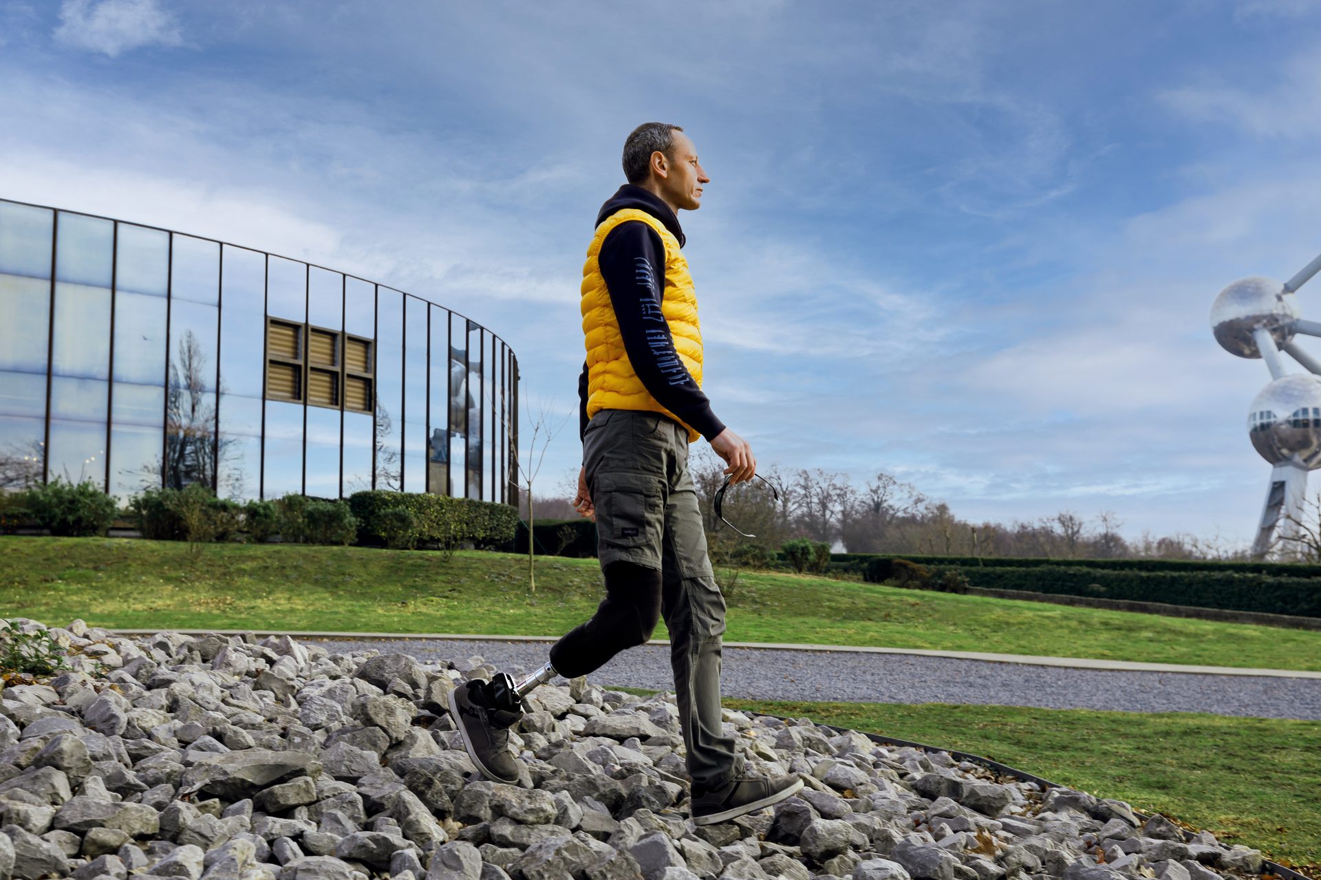
Source: Axiles Bionics, “Transtibial amputee with Lunaris foot prosthesis” — CC BY-SA 2.0. https://commons.wikimedia.org/wiki/File:Transtibial_amputee_with_Lunaris_foot_prosthesis.jpg
The patellar tendon bearing (PTB) socket, introduced by Radcliffe at the University of California in the 1950s, dominated transtibial design for decades and remains a foundational board concept. Its defining feature is selective load distribution: pressure-tolerant areas are loaded and pressure-sensitive areas are relieved through cast rectification. On the positive plaster model, add plaster to relieve, remove plaster to load. Adding plaster creates a relief (concavity) in the socket; removing creates a buildup (convexity). The patellar tendon is the primary weight-bearing surface, loaded by the PTB bar built into the anterior wall. The socket is set at approximately 5 degrees of initial flexion, which loads the patellar tendon, facilitates quadriceps contraction, resists genu recurvatum, and accommodates mild contractures.
Three PTB variants address short or unstable limbs. The standard PTB (mid-patellar trim line) is for standard-length limbs with a stable knee and is least restrictive. The PTB-SC (supracondylar) extends above the femoral condyles medially and laterally for self-suspension and ML stability with short limbs. The PTB-SC/SP (supracondylar/suprapatellar) extends above the condyles and patella, providing maximum ML+AP stability for very short limbs and ligamentous knee instability; it is the most restrictive of knee ROM and prevents genu recurvatum via the high anterior wall.
The total surface bearing (TSB) socket is the modern evolution: pressure is distributed uniformly across the entire limb through a gel liner (silicone, urethane, or thermoplastic elastomer) over a minimally rectified cast. Total contact is essential because air gaps cause pistoning and shear. The liner rolls onto the limb first, then the limb-and-liner enters the rigid outer socket. Suspension is often integrated into the liner (pin, lanyard, or suction seal) rather than relying on socket shape.
A systematic review found no overall superiority of either design, but TSB sockets had significant advantages on stairs and inclines and were associated with greater activity in active and traumatic amputees. The modern trend is strongly toward TSB with gel liners; the boards heavily test PTB principles because the underlying pressure-tolerant-versus-sensitive anatomy applies to both.
High Yield — Residual limb and foundational sockets
- Energy cost transtibial traumatic ~10-25%; vascular ~40%; bilateral transtibial ~40% traumatic / 60-80% vascular. Transfemoral traumatic ~60-70%; vascular ~100%. Knee preservation matters.
- Ideal residual limb: cylindrical or conical; tibia transected within proximal 50% (middle third most common); fibula 1-2 cm shorter than tibia; tibial crest beveled; myodesis preferred over myoplasty; posterior or posterolateral scar.
- Knee flexion contracture is the most common and most preventable post-amputation complication; >15 degrees is very difficult to accommodate.
- PTB socket (Radcliffe, 1950s) = patellar tendon as primary weight-bearing surface; 5 degrees initial flexion; rectification (add plaster to relieve, remove plaster to load).
- PTB variants: PTB-SC (supracondylar; short limb, ML stability); PTB-SC/SP (supracondylar/suprapatellar; very short limb + ligamentous knee instability; max ML + AP control).
- TSB socket = uniform pressure distribution via gel liner; minimally rectified cast; modern standard for active patients; statistically significant advantages on stairs and inclines.
Mnemonic — Add to relieve, remove to load (rectification rule)
On the positive plaster model: Add plaster where you want to Absolve the limb of pressure (creates relief/concavity in the socket); Remove plaster where you want to Root the load into a tolerant area (creates buildup/convexity in the socket). The model and the socket are inverses: every gap on the model becomes a bump in the socket, and every bump on the model becomes a gap in the socket. Once you internalize this inversion, every PTB pressure-relief and pressure-loading question becomes mechanical.
Every single step your patient takes relies on that foundational anatomical pressure map, and the most expensive advanced components in the world cannot compensate for a nerve trapped against a poorly relieved fibular head.
— PO-04 podcast, ~1:01:44