Before You Listen
- Prerequisites: Glasgow Coma Scale (GCS) severity stratification; basic ventricular and cisternal cerebrospinal fluid (CSF) anatomy; the difference between upper motor neuron (UMN) and lower motor neuron (LMN) signs; Modified Ashworth Scale (MAS) for spasticity.
- Runtime: 1 hour 21 minutes.
- Topic in one line: post-traumatic seizures (PTS) and the Temkin 7-day rule, post-traumatic hydrocephalus and the Hakim-Adams triad, the cranial nerve (CN) 1-7-8 ranking, convergence insufficiency at the 5 cm vestibular ocular motor screening (VOMS) threshold, heterotopic ossification (HO) at the hip with alkaline phosphatase as the earliest marker, venous thromboembolism (VTE) prophylaxis sequencing, paroxysmal sympathetic hyperactivity (PSH), post-traumatic headache, sleep architecture failure, post-traumatic hypopituitarism with the Ghigo screening algorithm, neurogenic bladder/bowel patterns specific to TBI, and pressure injury risk on the Braden Scale with staging by the NPIAP stages.
Vignette. A 58-year-old man is 8 weeks out from a severe TBI sustained in a fall from a roof. During inpatient rehabilitation, the nursing team reports that his right hip will not flex past 70 degrees during morning stretches, and the periarticular tissue feels warm and firm. A serum alkaline phosphatase drawn that morning is 312 U/L (baseline 95). Plain radiographs of the hip are unremarkable. He has been on enoxaparin since hospital day 3 and his last venous duplex was negative one week ago.
What is the most likely diagnosis, the single most sensitive imaging study to confirm it, the prevention strategy that should already have been in place, and the criteria that govern the timing of definitive surgical excision?
Section 1: Post-Traumatic Seizures and the Seven-Day Rule
Bottom line: post-traumatic seizures (PTS) split into immediate (<24 h), early (24 h–7 d), and late (>7 d) categories with different mechanisms; antiepileptic prophylaxis works for the first 7 days only and does nothing for late epilepsy; penetrating injury is the highest-risk profile at 33–50%.
Post-traumatic seizures are seizures that occur as a direct consequence of traumatic brain injury (TBI), and the temporal classification is not arbitrary. Each window reflects a distinct pathophysiology that drives a distinct treatment. Immediate seizures occur within 24 hours of impact and represent direct mechanical depolarization, ionic flux, and glutamate excitotoxicity. They are generally benign impact phenomena and do not predict long-term epilepsy. Early seizures occur between 24 hours and 7 days and result from acute cascades, including cerebral edema, intracranial hemorrhage expansion, blood-brain barrier disruption, and inflammatory mediator release. They drive intracranial pressure and metabolic demand, both of which compound secondary brain injury. Late seizures occur after 7 days and result from chronic processes, including cortical reorganization, gliotic scarring, aberrant network formation through epileptogenesis, iron deposition from hemoglobin degradation, and chronic neuroinflammation. When late seizures recur, the diagnosis becomes post-traumatic epilepsy (PTE). Focal seizures, often focal aware, predominate. Most post-traumatic seizures begin at 1–3 months after injury, 50–66% by one year and 75–80% by two years. After a first late seizure, recurrence reaches about 86% within two years.
The 7-day boundary marks the point at which acute provoked seizures (suppressible by drugs) give way to structural epileptogenesis (which current pharmacology cannot stop). Prophylaxis works for the acute window and only for the acute window.
Incidence scales sharply with severity. Mild TBI carries a 1–1.5% overall seizure incidence with <1% late risk. Moderate TBI runs ~2.9%. Severe blunt TBI rises to 12–17% overall with 10–17% late risk. Penetrating TBI sits at 33–50%, the highest of any category, and the risk persists for decades, with elevated risk extending 15–35 years after injury. A separate CT-confirmed combat TBI cohort (66 followed) showed late seizures in 25.8% at a median 7.4 years; that figure describes the whole combat cohort, not penetrating injury specifically.
Board answer vs. current evidence — Mild TBI carries a low late seizure risk, classically under 1% — this is likely the board-tested answer, but more recent evidence indicates post-traumatic epilepsy after mild TBI may reach 4.5% at 5 years in military populations, roughly tripling baseline epilepsy risk, so the classic <1% figure likely understates risk in certain cohorts. Answer the board-canonical version on the test.
Risk factors for late PTS include penetrating injury (33–50%), intracranial hematoma whether epidural, subdural, or intraparenchymal (25–30%, driven by iron from hemoglobin breakdown), early seizure within the first week (~25%), depressed skull fracture (3–70% depending on depth and dural penetration), prolonged coma or post-traumatic amnesia >24 hours (up to 35%), and cortical contusion of temporal or frontal cortex. GCS <10, age >65, craniotomy, and multiple hemorrhage types each independently raise risk. Among lesion types, bilateral parietal contusions (about 66%) and dural penetration (about 63%) carry the highest late-seizure risk. Midline shift over 5 mm is a further marker.
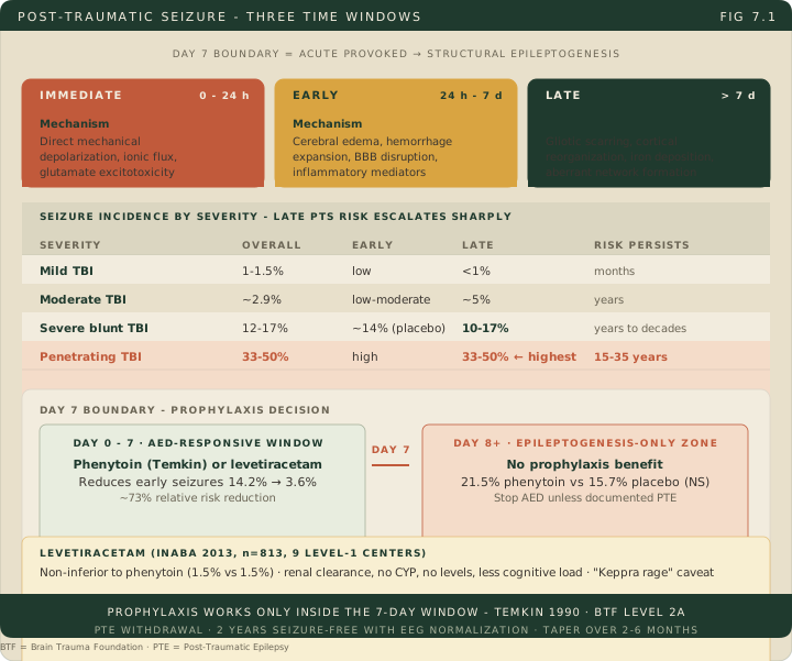
The evidence base rests on the Temkin trial, a randomized double-blind placebo-controlled study published in the New England Journal of Medicine in 1990 that enrolled 404 patients with serious head trauma. Patients received intravenous phenytoin loading within 24 hours of injury, followed by oral maintenance. Within the first 7 days, the phenytoin arm had a 3.6% early seizure incidence compared to 14.2% with placebo, a ~73% relative risk reduction. Beyond 7 days, late seizures occurred in 21.5% on phenytoin versus 15.7% on placebo, with no statistically significant difference and a numerical trend favoring placebo at 2 years. The Brain Trauma Foundation (BTF) codified this as a Level 2A recommendation: phenytoin (or equivalent) for 7 days, then stop.
Levetiracetam has largely displaced phenytoin in practice. The Inaba 2013 prospective comparison at two Level I trauma centers (n=813, 406 versus 407) found early seizures in 1.5% of each group, a nonsignificant difference. Levetiracetam is renally cleared (no cytochrome P450 entanglements), needs no drug-level monitoring, and produces less cognitive impairment. Its main caveat is behavioral irritability (“Keppra rage”), which is a real liability in a population already prone to agitation. A 2024 survey of 84 responding US trauma centers found levetiracetam in use at 82 of 84 (about 98%), although BTF technically still lists phenytoin in the Level 2A recommendation because that was the molecule used in Temkin.
For patients who develop documented PTE requiring chronic anticonvulsant therapy, the generally accepted minimum for considering medication withdrawal is 2 years seizure-free; an abnormal EEG raises recurrence risk and informs the decision, and the taper is gradual. Drug-resistant focal PTE is referred for epilepsy surgery evaluation, where resection and vagus nerve stimulation are the options.
High Yield — Post-traumatic seizures
- Temkin 7-day rule: prophylaxis prevents early (24 h–7 d) seizures, not late epilepsy. Stop at day 7 when no seizure has occurred; a treated early seizure or documented PTE follows the treatment protocol instead.
- Penetrating TBI = 33–50% late seizure risk, the highest category. Risk persists 15–35 years.
- Levetiracetam has practically replaced phenytoin: no CYP interactions, no level monitoring, less cognitive load. Watch for behavioral irritability.
- Iron from hemoglobin breakdown is directly epileptogenic, which explains the 25–30% late risk after intracranial hematoma.
- PTE diagnosis: recurrent unprovoked late seizures (>7 days post-injury); under the ILAE definition a single late seizure qualifies only when the estimated ten-year recurrence risk is at least 60 percent.
- First late seizure: recurrence reaches about 86% within two years.
- Semiology and onset: focal (often focal aware) seizures predominate; most PTS begin at 1–3 months, 50–66% by one year, 75–80% by two years.
- Highest-risk lesions: bilateral parietal contusions (about 66%) and dural penetration (about 63%); midline shift >5 mm is a further marker.
- Drug-resistant focal PTE is referred for epilepsy surgery evaluation (resection or vagus nerve stimulation).
Board Trap — Continuing prophylaxis past day 7
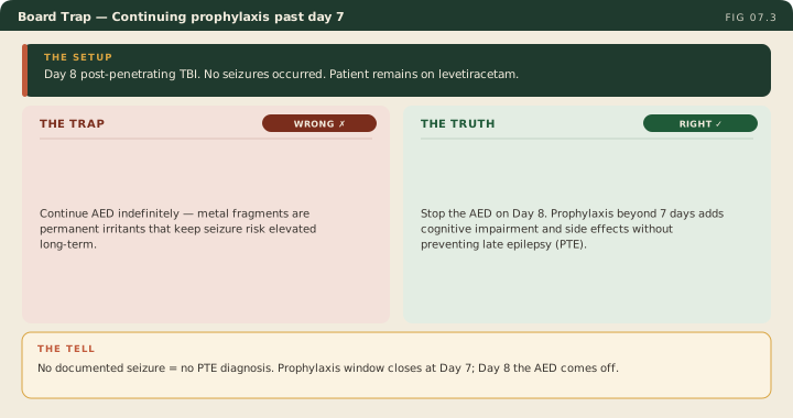
The reflex to “keep them on it just in case” is wrong. Continuing phenytoin or levetiracetam beyond 7 days adds cognitive impairment, drug interactions, and gingival hyperplasia (phenytoin) or irritability (levetiracetam) without preventing late epilepsy. Day 8 is when the AED comes off when no seizure has occurred. A patient treated for an early seizure continues antiseizure medication by the treatment protocol rather than the prophylaxis clock, and documented PTE means chronic therapy.
The medication simply suppresses the acute electrical irritability. It does not alter the fundamental structural pathology of the scarring.
— TBI-07 podcast, ~12:13