Before You Listen
Episode Setup
- Topic in one line: the procedural toolkit of modern physiatric pain medicine, anchored in the three epidural steroid injection (ESI) approaches (interlaminar, transforaminal, caudal), the particulate-versus-non-particulate steroid rule for cervical transforaminal injections, facet joint pain diagnostic algorithm (medial branch block to radiofrequency ablation (RFA)), sacroiliac joint procedures, vertebroplasty and kyphoplasty, neuromodulation (spinal cord stimulation (SCS), dorsal root ganglion (DRG) stimulation, peripheral nerve stimulation), intrathecal drug delivery (morphine and ziconotide), botulinum toxin for chronic migraine and myofascial pain, ketamine infusion for refractory complex regional pain syndrome (CRPS), and the radiation safety principles (inverse square law, As Low As Reasonably Achievable (ALARA)) that govern fluoroscopic procedures.
- Prerequisites: REHAB-01 (gate control theory, dorsal horn anatomy, opioid receptors and pharmacology) and REHAB-02 (CRPS Budapest criteria, sympathetic block anatomy by region, cancer pain ladder).
- Runtime: 53 minutes.
Vignette. A 64-year-old woman with osteoporosis is referred for axial low back pain following a fall onto her buttocks 5 days ago. She was admitted for pain control; despite scheduled analgesics including opioids and a thoracolumbar orthosis, her pain remains 9 out of 10, worse with sitting and weight bearing, and she cannot sit or stand for therapy. She has no radicular symptoms. She has no incontinence and no lower-extremity weakness. Magnetic resonance imaging (MRI) of the lumbar spine shows an acute T2-hyperintense fracture at L1 with anterior wedging of approximately 30 percent and bone marrow edema; there is no retropulsion, no spinal canal compromise, and no posterior wall involvement. She is also being evaluated separately for chronic axial low back pain at L4-L5 and L5-S1 with 80 percent or greater relief from each of two medial branch blocks performed on separate days using lidocaine and bupivacaine. Vitamin D is 18 ng/mL.
What procedure addresses the acute fracture, what is the anatomic rule for the radiofrequency ablation that follows the dual comparative blocks for her chronic pain, what nerve targets are required to denervate the L4-L5 and L5-S1 facet joints, and what radiation safety principle governs operator positioning during the fluoroscopic portions of these procedures?
(Answer at the end of this chapter)
Section 1: Epidural Steroid Injections — Three Approaches and the Particulate Steroid Rule
Bottom line: ESIs treat radicular pain from disc herniation or foraminal stenosis through three approaches: interlaminar (loss-of-resistance through the ligamentum flavum with a Tuohy needle, posterior epidural space, lower vascular risk), transforaminal (fluoroscopy-guided needle placement in the safe triangle of the Scotty dog view at the inferior pedicle, anterior epidural space, most precise but highest vascular risk), and caudal (through the sacral hiatus between the sacral cornua, lowest dural puncture risk, requires larger volume); the absolute rule for cervical transforaminal ESI is to use non-particulate dexamethasone because particulate methylprednisolone, triamcinolone or betamethasone can embolize into the vertebral artery or radicular feeders to the anterior spinal artery and cause cord infarction or posterior circulation stroke; contrast under live fluoroscopy confirms there is no vascular uptake before any steroid is injected.
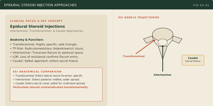
The primary indication for an epidural steroid injection (ESI) is radicular pain from disc herniation or foraminal stenosis, where steroid reduces inflammation around the compressed nerve root. Three anatomic approaches exist, each with characteristic indications, technique, and risks.
The interlaminar approach is the most traditional. The needle traverses the skin, subcutaneous tissue, supraspinous and interspinous ligaments, and the ligamentum flavum into the posterior epidural space. The hallmark technical maneuver is loss of resistance: a glass syringe filled with saline or air is attached to a Tuohy needle, and continuous gentle pressure on the plunger is applied while advancing. As long as the needle tip is embedded in dense ligamentum flavum, the plunger does not move; once the tip penetrates the ligamentum flavum into the epidural space, resistance disappears and the plunger glides forward freely. This loss confirms entry. The interlaminar approach delivers medication to the posterior epidural space, requiring diffusion anteriorly to reach the nerve root.
In the cervical spine the interlaminar approach carries its own level rule, and it is the companion to the transforaminal particulate-steroid rule above. Cervical interlaminar epidural injections should be performed at C7-T1 (or C6-C7 if necessary), and per the 2015 Multi-Society Pain Workgroup consensus should not be performed above C6-C7. The reason is dimensional: the posterior epidural space narrows progressively as you ascend the cervical spine, measuring only a millimeter or two at mid-cervical levels with the neck flexed, so there is almost no margin between the ligamentum flavum and the cord. The ligamentum flavum is also frequently midline-incomplete in the cervical spine, which removes the tactile loss-of-resistance endpoint the technique depends on. Injectate placed low reaches the target levels by spreading cephalad, so entering at C7-T1 costs little and avoids operating where the cord is closest to the needle. It is technically simpler than transforaminal injection and carries lower risk of vascular injection.
Fluoroscopic technique for interlaminar injections. The patient lies prone, and the AP view selects the interlaminar space: a Tuohy needle enters at the lower margin of the interlaminar opening, in the midline or paramedian (parasagittal), angled toward the pedicle on its own side. The paramedian needle goes on the side of the symptoms; when that side has no open path, it goes on the side that has one. A paramedian needle delivers the drug forward and to its own side: parasagittal interlaminar contrast reached the anterior epidural space in all 29 patients, against 75 percent of transforaminal injections, and in a 12-patient series the spread was unilateral in every case. The AP view shows no depth, so depth is checked in a contralateral oblique (CLO) view: the C-arm rotates away from the needle side, 45 degrees in the lumbar spine and 50 degrees at the cervicothoracic junction, the laminae appear as ellipses, and the ventral interlaminar line (VILL), the line joining the ventral margins of the laminae, marks the posterior boundary of the epidural space. The needle is brought to the VILL under CLO, and loss of resistance carries it the last millimeters; in the cervical spine resistance is lost at or within 2 mm of the VILL, and the needle goes no more than 2.5 mm past it. The lateral view is the weaker depth view: the tip is poorly seen, and lumbar epidural contrast touched the ventral laminar margin on CLO in 100 percent of patients but the spinolaminar junction on lateral in only 29 percent. A false loss of resistance occurs superficial to the epidural space, on 53 percent of first cervical attempts in one series; contrast posterior to the VILL on CLO is non-epidural even when the AP epidurogram looks epidural, so the AP view alone never confirms placement. Every interlaminar injection uses AP plus a lateral or CLO view and a test dose of contrast. Intrathecal contrast shows no dense band along the VILL and a homogeneous column within the canal, and intradural contrast forms a globular collection or a tram track; either means stop, with no local anesthetic and no steroid. A vascular pattern means abort. Intrathecal injection is more common with interlaminar than with transforaminal access, from deep placement, a midline gap or frailty in the ligamentum flavum, and central stenosis; the target is the space between the ligamentum flavum and the dura. In 10,000 fluoroscopic epidurals, dural puncture occurred in 0.8 percent of lumbar interlaminar injections, and intravascular entry in 0.5 percent, against 7.9 percent for lumbar transforaminal injections. A postdural puncture headache begins within 5 days and eases lying flat; an epidural blood patch treats a headache that fails conservative care. In the cervical spine, prior imaging must show enough epidural space at the target level before an interlaminar injection, because the epidural space is widest at C6-T1 and ligamentum flavum gaps increase at each higher level. The transforaminal endpoint is a bony target confirmed on lateral; the interlaminar endpoint is tactile, and the CLO view is its check.
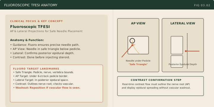
The transforaminal approach is fluoroscopy-guided and delivers medication directly to the anterior epidural space at the targeted nerve root. The needle enters through the neural foramen, placing the steroid directly at the disc-nerve interface. The fluoroscopic landmark is the Scotty dog view, an oblique view of the lumbar spine in which the posterior elements form a profile resembling a Scottish terrier: the pedicle is the eye, the transverse process is the nose, the superior articular process is the ear, the pars interarticularis is the neck, the inferior articular process is the front leg, and the lamina is the body. A pars fracture (spondylolysis) appears as a collar across the dog’s neck. The needle is placed in the safe triangle: the region bounded by the inferior border of the pedicle above, the lateral border of the vertebral body medially, and the exiting nerve root laterally and below. An alternative target is the Kambin triangle, the posterolateral safe zone of the foramen bounded by the exiting nerve root superiorly, the traversing nerve root medially, and the superior endplate of the lower vertebra inferiorly.
Fluoroscopic technique. Imaging starts in AP, with the C-arm tilted until the target endplate is squared (the beam tangential to the inferior endplate of the upper vertebra). The C-arm then rotates to an ipsilateral oblique until the Scotty dog appears with an unobstructed path to the target; the angle is set by level and anatomy, about 25 degrees in one technique guide and 30 to 45 degrees in one clinical series. The needle advances coaxially, “down the beam”, so the hub projects as a dot over the target. In the supraneural (subpedicular, safe triangle) approach, the oblique target is just below the eye at 6 o’clock (shifted slightly lateral, about 5:30 for a right-sided injection); on AP the tip sits just under the pedicle at or just lateral to 6 o’clock and stays lateral to the midpoint of the pedicle, because a more medial tip enters the central canal and risks intradural or intrathecal injection; on lateral it advances about halfway across the pedicle and stays in the posterior half of the foramen, just below the pedicle, because the vessels lie in the anterior half. The tip stays in the foramen; the injectate reaches the anterior epidural space. In the infraneural (Kambin, retrodiscal) approach, the oblique target is the junction of the superior articular process and the superior endplate of the lower vertebra; on AP the tip stays lateral to the mid-pedicular line to avoid dural puncture; on lateral it sits in the inferior foramen and does not enter the disc. The infraneural approach exists because of the arteries: thoracolumbar radicular arteries that feed an anterior radiculomedullary artery lie in the anterosuperior quadrant of the foramen in 96 percent of cases, and the artery of Adamkiewicz lies in the superior half in 97 percent and was never found in the inferior fifth. Its trade-off is the disc: inadvertent intradiscal injection occurred in 4.7 percent of 257 retrodiscal injections versus 0.25 percent of subpedicular injections. Before any steroid, contrast (0.5 to 1 mL) goes in through extension tubing under live fluoroscopy in the AP view, with digital subtraction when available. The desired pattern outlines the exiting spinal nerve and flows into the epidural space medial to the pedicle, reaching the medial aspect of the superior pedicle, so the expected AP picture is contrast tracking medially and cephalad around the pedicle. Ventral flow reaches the anterior epidural space: a superior-anterior tip produced ventral epidural flow in 100 percent of injections and a superior-posterior tip in 61.4 percent, and the superior-posterior tip produced dorsal epidural flow in 95.5 percent, so dorsal epidural contrast on the lateral view is the expected finding from a posterior-half tip, not an error. Contrast that stays lateral, along the root with no medial epidural flow, fails to show the desired pattern and does not by itself locate the tip: in one series this root-only pattern occurred with 7 of 12 extraforaminal tips and with 8 of 86 foraminal tips, so the tip position is reassessed in more than one view, the needle adjusted, and contrast repeated before any injectate. A venous pattern means reposition, by advancing the needle, and repeat contrast. An arterial pattern is fleeting and lies away from the target: in one lumbar TFESI it ran as a central, longitudinal line along the anterior spinal artery several levels away while the epidural pattern at the target looked correct, and it was detected on live fluoroscopy; an arterial pattern means abort. An intrathecal injection gives a myelographic, homogeneous contrast column within the canal, and an intradural injection gives a globular collection or a tram-track of two parallel lines; for transforaminal injections the AP view is the most critical view for recognizing both. Either pattern means abort, with no local anesthetic and no steroid, because intrathecal local anesthetic produces spinal anesthesia, which at lumbar levels is a motor and sensory block of the lower extremities. A local anesthetic test dose (1 to 2 mL of preservative-free lidocaine) follows, then non-particulate dexamethasone.
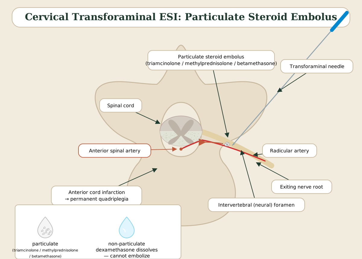
The critical safety distinction for cervical transforaminal ESI is steroid choice. Particulate steroids (methylprednisolone / Depo-Medrol, triamcinolone / Kenalog, betamethasone / Celestone Soluspan) are crystalline suspensions whose particles can embolize if inadvertently injected into a radicular artery. In the cervical spine, the vertebral artery and radicular feeders to the anterior spinal artery course through or adjacent to the neural foramen near the needle path. Particulate steroid embolization can produce catastrophic spinal cord infarction, posterior circulation stroke, or death. Non-particulate dexamethasone is required for cervical transforaminal ESI (a true solution, not a suspension); betamethasone, including the branded Celestone Soluspan formulation (a phosphate plus acetate mixture), is a particulate suspension and carries the same embolic risk as methylprednisolone and triamcinolone. This rule is absolute. In the lumbar spine the catastrophic risk is lower (the artery of Adamkiewicz arises on the left in about three quarters of people, from an intercostal or upper lumbar segmental artery between T9 and L1), but many practitioners now use non-particulate agents universally.
The caudal approach enters the epidural space through the sacral hiatus, the opening at the inferior end of the sacrum where the laminae of S5 are absent. The hiatus is identified by palpating the sacral cornua flanking it. The needle is directed superiorly at approximately 45 degrees through the sacrococcygeal ligament into the caudal epidural space. The primary advantage is the lowest risk of dural puncture because the thecal sac typically terminates at S2, well above the hiatus entry point, which makes the caudal approach useful when spinal headache risk is high. It does not lower bleeding risk: epidural hematoma arises from the epidural venous plexus whatever the route, so anticoagulants and antiplatelets are managed per ASRA pain-procedure guidance as for any epidural injection. The disadvantages are the larger required volume (often 10 to 20 mL) and less precise delivery, since medication must travel upward through the epidural space.
High Yield — ESIs
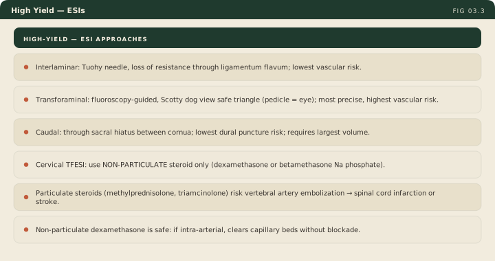
- Interlaminar: Tuohy needle through ligamentum flavum, loss of resistance = entry; posterior epidural space; lowest vascular risk.
- Transforaminal: fluoroscopy-guided, safe triangle of Scotty dog view (pedicle = eye); most precise; highest vascular risk.
- Caudal: through sacral hiatus between cornua; lowest dural puncture risk; requires larger volume.
- Cervical transforaminal ESI: must use non-particulate dexamethasone. Particulate methylprednisolone / triamcinolone / betamethasone (Celestone Soluspan) risk vertebral artery embolization → spinal cord infarction or stroke. Absolute rule.
The interlaminar approach relies on the tactile loss of resistance through the dense ligamentum flavum to enter the posterior space, offering the lowest vascular risk. The transforaminal approach uses live fluoroscopy to target the safe triangle of the scotty dog for the anterior space, offering the absolute most precision but carrying the highest vascular risk. And the caudal approach. Right. The caudal approach enters incredibly low through the sacral hiatus, safely carrying the lowest dural puncture risk.
— REHAB-03-a podcast · ▶ Listen from 55:57